 |
| |||||||||||||||||||||||||||||||||||||||||||||||||||||||||||||||||||||||||||||||||||||||||||||||
- for curative programs in rural hospitals, dispensaries and
refugee camps
- for the use of doctors and nurses
1993 - THIRD EDITION
NOTE FROM THE CD-ROM EDITORS: THIS MANUAL SHOULD BE USED BY MEDICALLY TRAINED PERSONS ONLY. THE GREATEST CARE HAS BEEN GIVEN TO ACCURATE REPORT BUT IT CAN NOT BE TOTALLY EXCLUDED SOMETIMES A TYPESETTING OR SCANNING ERROR HAS OCCURED (ON AVERAGE 1 OUT OF 2000 CHARACTERS IN TEXT AND 1 OUT OF 200 to 300 DIGITS IN TABLES).
DOSES OR MEDICAL ACTIONS MENTIONED HERE SHOULD BE CHECKED WITH THE COMMON MEDICAL SCIENTIFIC AND PHARMACEUTICAL KNOWLEDGE AND WITH THE ACTUAL LOCAL SITUATION AND PARTICULARITIES AS ASSESSED AND JUDGED BY A MEDICALLY TRAINED PERSON.
|
| |||||||||||||||||||||||||||||||||||||||||||||||||||||||||||||||||||||||||||||||||||||||||||||||
Acknowledgments
Clinical guidelines
Diagnostic and treatment manual
EDITOR:
J.C. DESENCLOS (MD)
THIRD EDITION COORDINATED BY
P. BIBERSON (MD)
J. RIGAL
(MD)
Contributions (in alphabetical order):
H. Audrain (N), P. Autier (MD), M.J. de Chazelles (Oph), J. Combreau (MW), E. Couturier (MD), B. Faucher (MD), V. Fauveau (MD), L. Flachet (MD), A. Fourrier (Ph), J.J. Frere (MD), D. Jorland (CD), J. Lagoutte (MD), A. Moren (MD), B. Moriniere (MD), F. Mounis (O), B. Pecoul (MD), J. Pinel (Ph), M. Postel (MD), B. Renchon (Ph), V. Schwoebel (MD), C. Segala (MD), P. Sotias (MD), V. Van Stirtegheim (MD).
FRENCH-ENGLISH TRANSLATION COORDINATED BY
P. HAKEWILL (MD)
J. PORTER (MD)
R. KESSEL (MD)
(MD) Medical doctor, (MW) Midwife, (N) Nurse, (O) Osteopath, (Ph) Pharmacist, (D) Dentist, (Opht) Ophthalmologist
We would like to thank Dr S. Sorensen (Essential drugs program, WHO), Professor M. Rey (WHO and Dr Houlemarre (Centre International de l'Enfance) for their advice and suggestions; Dr G. Desve, Mr M. Pottier and Miss O. Hardy for their technical assistance in the writing or this manual.
This book would not have been possible without Ms Evelyne LAISSU who was responsible for the design and layout.
|
| |||||||||||||||||||||||||||||||||||||||||||||||||||||||||||||||||||||||||||||||||||||||||||||||
Foreword
This clinical manual is a collective work, for daily field practice.
We have tried to incorporate information from various sources:
the field experience of Medecins sans Frontieres personnel, the recommendations
from reference institutions such as World Health
Organization (W.H.O.) and
from text books and monographs most relevant to the domain of medical care in
developing countries (see bibliography).
This manual is for doctors, nurses and other health professionals responsible for curative care in rural dispensaries and hospitals, as well as in displace people or refugee camps.
It covers the curative and to a lesser extent the preventive aspects of the main conditions encountered in the field. It should function as a supportive tool towards the elaboration of an adapted health policy. The introduction of this manual will emphasize the basis of such a policy.
With a view to future revisions and to keep the work as close as possible to field realities, the authors would be grateful for critical comments and suggestions from users of this manual.
Comments should be send to
Medecins sans Frontieres - Service medical
8 rue Saint-Sabin
- 75544 Paris Cedex 11 - France
Tel.: (33.1) 40.21.29.29 - Fax: (33.V
48.06.68.68 - Tlx: 214 360
F
|
| |||||||||||||||||||||||||||||||||||||||||||||||||||||||||||||||||||||||||||||||||||||||||||||||
How to use these guidelines
Organisation
The information you are looking for can be found:
1. At the beginning of the manual in the table of contents: numbers of chapters with page numbers.
2. At the end of the manual in the alphabetical index whith the lists all diseases
Abbreviations used
mg = milligramme
g = gramme
kg= kilogramme
d = day
x
= times
stat = at once; one single dose
AFB = acid fast bacilli
BP =
blood pressure
CCF = congestive cardiac failure
CSF = cerebrospinal
fluid
GlT = gastro-intestinal tract
Hct = haematocrit
MCH =
maternal-child health
0RS = oral rehydration salts
0RT = oral rehydration
therapy
PID = pelvic inflammatory disease
PO = per os (orally)
IM =
intramuscular
IV = intravenous
SC = subcutaneous
IU = international
units
MIU = million international units
PR = per rectum
PV = per
vaginam
PUO = pyrexia of unknown origin
RBC = red blood cell
RR =
respiratory rate
RTI = respiratory tract infection
spp = species
STD =
sexually transmitted diseases
TB = Tuberculosis
WBC = white blood cell
- Cotrimoxazole = mixture of sulfamethoxazole (SMX) +
Trimetoprim (TMP)
Usual dosage is: 400 mg SMX + 80 mg TMP
- Peni G = Benzyl penicillin = Crystalline penicillin G
- PPF = Fortified procaine penicillin = mixture of procain benzyl penicillin and Benzyl penicillin
Conversion °C into °F: remove 2, multiply by 2, add
30
Conversion °F into °C: remove 30, divide by 2, add 2
International non proprietary name for drugs
The International Non-proprietary Name (INN) of drugs is used in this manual. A list of equivalent commercial running name can be found.
|
| ||||||||||||||||||||||||||||||||||||||||
Clinical Guidelines and Treatment Manual (MSF, 1993, 319 p.)
Introduction
In a health program adapted to the needs of a developing country, the curative care is an important component. Initially however, more important measures have to be implemented to provide the foundations for all programs aimed at improving the health of a community. These measures are related to:
- sanitation,
- nutrition,
- hygiene,
- immunization,
- maternal and child health,
- health education,
- health workers training,
- community awareness and participation.
These measures should interact and complement the curative care component of the health program.

Figure
1
Objectives for a curative health program
At the individual level
The objective is to cure the patient and to minimize or prevent the consequences of illness (eg. transmission).
At the community level
The objectives are to reduce the mortality and morbidity attributable to the common severe illnesses in the community.
For a few infectious endemic diseases
Curative care can reduce transmission of certain diseases (e.g. TB, leprosy, trypanosomiasis, bilharzia) provided a high proportion of the infected community is treated.
Strategy
In developing countries there are enormous needs and limited resources. The resources should be aimed at the diseases, amenable to effective treatment in the field, which are causing high mortality and morbidity (priority diseases).
Priority diseases can vary from one geographical region to another, but a standard epidemiological profile remains. In order to get an accurate profile an initial assessment is necessary. It should be qualitative (descriptive), and if possible, quantitative (incidence, morbidity and mortality rates). This evaluation will characterise the most common diseases (e.g. diarrhoea, acute respiratory infections...) and will identify the exposed and high risk groups in the population (e.g. children < 5 years, pregnant women...). These diseases and high risk groups should be the targets of the program. This does not mean that curative care should be limited to these diseases and groups of people, but rather that the resources, particularly at the primary health care level, should be targeted at these groups.
In some instances (e.g. displaced or isolated persons) a complete evaluation is necessary. In other instances, such as a rehabilitation program or a study to reinforce an existing program, the Ministry of Health (MOH) may already have qualitative or quantitative data available and only a partial evaluation may be necessary.
The health care program can be defined and carried out as soon as priorities have been defined, and health policy and local resources identified (e.g. essential drug list, MOH management protocols, medical personnel and their training and the medical structure).
This manual, "Essential drugs - practical guidelines" and "Principales conduites a tenir en dispensaire" are additional tools to help evaluate, define and establish a health care program (e.g. management protocols, training, guidelines...).
Health care organization
In certain situations (e.g. displaced populations, refugees), a program has to be created, whereas in others, an existing program is evaluated so it can be improved.
Infrastructure and medical staff
Health centers, dispensaries, medical centers and hospitals are run by personnel with different skills and different levels of competence (e.g. community health workers (C.H.W.), medical auxiliaries, nurses, midwifes and doctors).
The evaluation should clarify their technical level. In refugee camps, most of the staff will have no previous training.
Medicines
Selection of medicines depends on the targets and needs identified in the epidemiological profile. However, are other restraints: cost, stability, administration route, duration of treatment and whether single or multiple drug doses are required.
The W.H.O. list of essential drugs (appendix 3), is the basic framework for establishing an essential drug list. A drug list should be defined in accordance with objectives, target diseases, epidemiological profile, medical staff competence and whether it is possible to refer severe cases. The quantitative and qualitative drug lists of the Emergency Health Kit (for 10.000 persons for 3 months) recommended by the W.H.O. and Medecins sans Frontieres are given as an example in appendix 4.
Drugs are listed under their Intemational Nonproprietary (generic) Names: INN.
Therapeutic protocols
These protocols are the foundation stone of any curative health program and should be standardised in order to have an effective impact on the target diseases.
The therapeutic protocols should:
- Give clear accurate instructions.
- Include the therapeutic uses and dosages of drugs, and the duration of treatment.
- Choose the most effective drug with least side effects.
- Be supported by epidemiological and clinical data and should be discussed and agreed by the users.
- Be practical, simple, understandable and adapted to the field.
- Encourage the training and retraining of medical staff.
- Encourage the organization of medical infrastructure (e.g. pharmacy, management...).
- Be periodically re-evaluated.
- Always use the national recommendations of the country.
The therapeutic protocols should be adapted to the skill and knowledge of the medical staff. They should cover: drug prescription, curative and preventive measures, cases which should be notified (e.g. epidemic threats: cholera, typhoid), and the grounds for referral to a superior level hospital.
Protocols should be adapted to:
1) The skill and knowledge of the medical staff
A doctor is trained in terms of diseases and syndromes (e.g. pneumonia, liver abscess) whereas a Community Health Worker (CHW) is trained in terms of symptoms (e.g. cough, fever). These two approaches are presented in Chapter 2 "Respiratory Diseases", with an introduction of the WHO program on respiratory conditions which is founded on a symptomatic approach.
2) The cultural milieu and environment
For example, if it is the custom to treat children with diarrhea with rice water, or for children with fevers to remain clothed, do not reprimand their parents.
3) The pharmaceutical supplies and local dosages of drugs
Dosages are often different between countries (e.g. chloroquine 100 mg or 150 mg tablets).
4) The improvement of patient treatment and compliance
It is recommended that prescribed treatments are short (< 5 days) and, if possible, in single or twice daily doses. "Stat dose" treatments, although less effective pharmacologically, do not rely on patient compliance (e.g. treat amoebiasis with a single dose of 8 metronidazole tablets (tab 250 mg) instead of a 7 day course). For the same reasons, the prescription should be limited to a maximum of 2 prescribed drugs. Injections should be avoided to reduce HIV transmission or B hepatitis.
Protocols should avoid classical mistakes like recommending the boiling of water when energy resources (e.g. wood) are limited.
Recommendations and examples of therapeutic protocols can be found in:
- The protocols from the "New Emergency Health Kit" (CHW level) to target diseases (see appendix 4).
- The clinical and treatment sections of this manual.
Diagnostic methods
These methods depend on the structure of the organization and on the technical expertise of the staff. Staff expertise directly influences protocol formulation and drug list contents.
As a rule, diagnosis is based on the clinical examination and basic laboratory investigations (as it is defined in WHO).
Clinical examination
The principles here described are for trained medical staff. The approach for the CHW is similar but simpler.
Quality history taking and clinical examination is vital. If poor, the process from syndrome etiology to diagnosis will likewise be poor, and the treatment inappropriate. It is important to master a technique of clinical assessment that is methodical, complete and rapid. A method is all the more necessary because in field conditions the laboratory support may be rudimentary and the practitioner may have to communicate with the patient via an interpreter.
The following examination framework should be adapted to conditions. It emphasizes the advantages of a methodical approach.
CIRCUMSTANCES OF THE EXAMINATION
- Routine, as in a MCH clinic for prenatal women and well babies. The emphasis of the examination will depend upon local circumstances eg prevalence of anemia, malnutrition.
- With respect to a complaint, the commonest of which tend to be pain, fever, cough, diarrhea, fatigue...
APPROACH TO HISTORY AND PHYSICAL EXAMINATION
- A methodical approach is vital. This will save time and reduce omissions.
- An interpreter will usually be necessary. He/she must have received prior training and there must be good rapport between the clinician and the interpreter. Eventually, a good interpreter takes a very active role in the clinical process and becomes far more than a simple translator. Choosing an interpreter requires thought; the person must be acceptable to the community and appropriate for the specific role (eg a woman for obstetrics and gynaecology).
- Learning the local words for major symptoms and diseases will allow the clinician to check that an interpreter, unfamilar to him or her (such as a relative), is giving an accurate rendition of the patient's complaints.
FRAMEWORK OF A CLINICAL ASSESSMENT
- History
· history of the present illness
· the
circumstances
· past history, family history
· current
medications, allergies
- Examination
The patient should be undressed if possible.
· General appearance: nutrition (weight and height of
children), hydration, temperature, pallor; does the patient look sick
?
· Examination by systems: respiratory, cardiovascular, etc. This part
of the examination in particular should be rigorously methodical.
- Laboratory Tests: if necessary.
- Diagnosis: This is a synthesis of all information gathered from the history, physical examination and laboratory tests. A diagnosis should be etiological but may of necessity be only symptomatic.
- Treatment
· etiological, ie treating the cause. This may have to
await the results of laboratory results;
· symptomatic;
· advice
to the patient, whether or not a treatment is given.
- All important clinical data should be recorded, either on a card or in a family health booklet. Especially note positive and significant negative clinical signs, laboratory results, and treatment given (generic name, dose, duration).
Role of the laboratory
A basic medical laboratory of the type described by WHO can play an important role. Nevertheless, there are special constraints upon the operation of a laboratory, which should not be underestimated. There are staff constraints (necessity of trained and competent technicians), logistic constraints (supply of reagents and other equipment), time constraints (a minimum of time is necessary for each examination) and quality constraints. If attention is not paid to the above considerations, the laboratory will loose its accuracy and therefore its useful purpose.
Two levels of examination should be considered:
BASIC EXAMINATION
- Stool exams direct and stained with Lugol's iodine solution, for parasites (ova, cysts, protozoa...).
- Blood slides: thick and thin smears (for malaria, trypanosomiasis, filiariasis, relapsing fever, screening for leucocytes): GIEMSA stain.
- Hemoglobin (Lovibond method).
- Urine exam:
· urine analysis: dipsticks for glucose and proteins.
- Sputum exam: Ziehl - Nielsen stain.
- Urethral and vaginal swabs: slides for gonococcus and trichomonas.
- CSF exam
COMPLEX EXAMINATIONS
Certain more complex examinations may be provided according to the specific program.
A laboratory can be used in two complementary ways:
- Clinically: examinations can be requested for individual patients according to the clinical picture. The aim will be to assist the practitioner in:
· diagnosis orientation (e.g. leucocytosis in blood
count);
· etiological diagnosis (e.g. stool exam for parasites, malaria
smear...).
- Epidemiologically: the aim will be to construct or to validate clinical and therapeutic protocols. One can investigate a sample of patients presenting with a particular clinical picture (symptoms and syndromes) specify the etiology of that clinical picture and thus arrive at an appropriate standardized therapeutic management protocol.
For example:
· Fever and chills: are they due to malaria ? Rather than being obliged to perform blood slides on every febrile patient, choose at random 100 patients presenting with these symptoms and investigate them. If a significant proportion of the blood slides are positive, such cases can henceforth be presumed to be malaria and treated according to an appropriate protocol.
· Bloody or mucusy diarrhea with no fever: the same approach can be used to determine if this clinical presentation is synonymous with amoebiasis and/or another intestinal parasite.
· This epidemiological method of using a laboratory is especially appropriate in responding to priority needs. It can be used in emergency or "normal" conditions. Bibliographical references n° 2 and 19 give two examples for malaria, one in a refugee camp, another one in Malawi.
The training
Training or retraining of medical staff should be directed at program objectives and means (e.g. target diseases, list of essential drugs, management protocols) and should take into consideration the technical level of the staff (to be evaluated). The training program should be defined according to local needs.
Community awareness and participation
It is necessary for curative care to cover the whole population and target diseases. Coverage should be as wide as possible.
For many reasons (e.g. ignorance, different cultural perception), a large proportion of severely ill patients may present late or may pass through the system without being cured. Coverage can be improved by increasing awareness, improving health education, encouraging the exchange of information at all levels and by improving the quality of care.
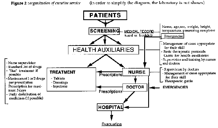
Management
Consider how to efficiently and effectively manage available resources. Figure 2 gives an example of organization of an out-patient department.
Evaluation
The evaluation of the common diseases and their effects on the community directly influence the nature of a program.
Program evaluation should be performed at the following levels:
- Level of functioning
Activity assessment, quantity of drugs used, prescription management, correct use of protocols, pharmacy management (orders, reports and stock keeping), all of this information should be used as indicators in program management. The morbidity rate at the dispensary level and its analysis is a useful epidemiological observation. Target disease variation in the community can be followed according to time, place, and population concerned (eg.: morbidity survey, appendix 2).
- Level of coverage
The aim is to determine what proportion of all patients affected by target diseases are reached by the program. Good coverage is an essential factor. The evaluation should be done on a representative sample of a target population (see below).
- Level of community impact
This aspect is difficult to evaluate. The evaluation relates to the objectives and should be expressed in terms of a decrease in morbidity and/or mortality. A mortality survey of a community can be conducted over a defined period of time. If the total population is known, a mortality rate can be determined.
Sample protocols for community surveys are available and have been used for evaluations (e.g. WHO, diarrheal disease program, but they require much organization and need to be repeated to give evidence of a trend).
Figure
2
|
| |||||||||||||||||||||||||||||||||||||||||||||
Clinical Guidelines and Treatment Manual (MSF, 1993, 319 p.)
Chapter 1 - A few symptoms and syndromes
Fatigue
Fatigue is one of the commonest presenting complaints. The term includes various subjective symptoms (lassitude, lack of energy etc), that are both physical and mental. In most of cases there is no pathological basis to be found, however it must not be forgotten that many diseases may present as fatigue. The symptom, as much as any other, requires a full, methodical clinical assessment.
Clinical features
The history and physical examination must define:
- Mode of onset: sudden or progressive, old or recent, isolated or associated with other symptoms, life situation (work, intense activity, recent illness, refugee displacement...).
- Nature of the fatigue: physical, intellectual, sexual...; whether it comes on in the morning (often psychosomatic) or evening (more usual).
- Any associated clinical features:
· Systemic features: anorexia, weight loss, fever, anaemia, all of which suggest a probable organic basis.
· Localizing features linked to a particular organ system, eg cough and haemoptysis in TB, dyspnea in cardiac failure or anaemia, abdominal pains in parasitoses, jaundice in hepatitis.
· Physical findings: the examination must be comprehensive:
· Nutritional status: weight (signs of recent loss), anaemia, signs of vitamin deficiency diseases...
· Cardiopulmonary: pulse, BP, chest auscultation...
· Abdomen: including liver, spleen...
· Lymph nodes
· Skin and mucus membranes
· Affect: anxiety, depression.
Diagnosis and Treatment (dispensary)
- If the fatigue is part of a syndrome, treat the cause.
- If there seems to be no organic basis, assume the complaint is
psychosomatic. Advise the patient to consult a traditional healer, who is
usually in a far better position to help. Depending on national recommendations,
a placebo may be prescribed, give:
multivitamins: 1 tab x 3/d x 5
days.
Pain
Pain is a common presenting symptom and of course may be caused by a range of conditions. Pain is a subjective experience. The same degree of pain will be expressed differently from patient to patient. There are also cultural differences. The assessment of the severity of pain in a given patient is thus difficult. The solution is to address the problem with a clinical approach that is both methodical and comprehensive.
Clinical features
The history of the pain elicited from the patient must define:
- Onset: sudden, subacute or progressive.
- Duration.
- Localization and radiation.
- Nature of the pain: colicky, burning, sharp, constricting, like a weight; and whether intermittent or continuous.
- Factors that induce or relieve the pain: posture, coughing, deep breaths, meals, specific foods, movement etc.
- Associated systemic features: fever, fatigue, weight loss, etc.
- Associated focal features: cough, diarrhea, vomiting, burning during micturition...
The physical examination should be oriented towards the organ system or region where the pain seems to be localized. The synthesis of the clinical data provides the diagnosis and orients therapy, both etiological and symptomatic.
Treatment
ETIOLOGICAL
That is, treatment of the cause of the pain.
SYMPTOMATIC (dispensary)
According to the nature of the pain.
- Headache
acetylsalicylic acid(PO): 3 g/d divided in 3 doses x 3-5 days
or
paracetamol (PO): 1.5 g/ d divided in 3 doses x 3-5 days
- Psychosomatic pains: consider this diagnosis if pains are multiple, fleeting, or shifting. Treat as for headache or refer to a traditional healer.
- Joint pains
acetylsallcyllc acid (PO):
Adult: 3 g/ d divided in 3 doses x 3-5 days
Child: 50 mg/kg/d divided in 3 doses x 3-5 days
- Inflammatory: tends to be worse at night. Look for an infectious cause (may require surgical drainage and antibiotics).
If acetylsalicylic acid is ineffective, treat
with:
indomethacin (PO):
Adult: 50 to 150 mg/d divided in 3 doses x 3-5 days
- Joint pain (especially monoarticular): exclude septic
arthritis.
Note that periarticular and bone pains with swelling and loss of
function of the limb may be due to scurvy: look for bleeding from the gums and
treat with:
Ascorbic acid (vitamin C) (PO):
Adult: 500 to 1,000 mg/d divided in 3 doses until recovery
Child: 100 to 300 mg/d divided in 3 doses until recovery
Give dietary Advice.
- Colic
· Gastrointestinal: exclude a parasitic infection. Do not
give acetylsalicylic acid (possibility of ulcer).
Depending on
severity:
N-butylhyoscine (PO):
Adult: 30-60 mg/d divided in 3 doses x 3-5 days
or
atropine (SC):
Adult: 0.5 to 1 mg by injection
Child: 0.01 to 0.02 mg/kg by injection
· Renal or biliary colic: same as above. If necessary:
noramidopyrine (IM or IV):
Adult: 500 mg by injection
- Very severe pain
noramidopyrine (IM or PO)
Adult: 500 mg as necessary
Or, if ineffective: pentazocine (IM or PO): 30 mg IM or 50 mg PO as necessary
Fever
Fever is common, and usually, related to an infection of viral, bacterial or parasitic origin. The type and duration of fever helps determine the diagnosis. Note that fever in the newborn has its own complications.
Fever may be defined as a rectal temperature above 37°C in the morning, and above 37°5C in the evening.The corresponding axillary temperature would be above 37°5C and 38°C. This definition is practical in hospital but not as satisfactory in a dispensary. Several factors have to be considered in taking a patient's temperature: the technique (axillary, oral, rectal), the quality of measurement, the patient compliance, and the time available. One usually considers that axillary temperature under estimates the core temperature by 0°5C.
- Clinically: any hyperthermia, even if it is only slightly above normal, could be significant (e.g. nocturnal febrile stage in tuberculosis). On the other hand, at dispensary and primary health care level, a higher threshold only should be considered (ea. axillary temperature > 38°C after 5 mins).
At hospital level, a finer thresh-holds can be adopted.
In all cases, it is essential to define these thresholds.
- Fever to be treated:
· In the infant and new born: over 38°C rectal temperature, and/or if there are signs of intolerance.
· In the adult: above 38°5C and/or if the patient is uncomfortable.
Clinical features
- The following complications can be brought about by fever in newborns and infants:
· Convulsions
· Dehydration
· Malignant
hyperthermia (collapse and coma)
They should be investigated and treated but moreover they should be prevented (see treatment).
- Clinical assessment is the main method of investigating the cause of fever. Epidemiological environment should also be considered. If available, a laboratory could be useful. The following guidelines are helpful. They should be adapted to the epidemiological context, level of medical staff and diagnostic methods.
FEVER AS A SERIOUS SYMPTOM OF INFECTION
- High fever, shivering, sweating, malaria endemic area (falciparum), headache, consciousness desorders (even minor) indicate severe malaria. Without treatment, it can cause death.Take a malaria smear and treat.
- High fever with general health impairement, with or without other signs indicates typhoid fever.
- High fever, stiffness and neurological signs indicate meningitis or meningocephalitis.
- High fever with:
· A hemorragical syndrome indicates meningococcemia, or hemorragic fever, or in an endemic area, relapsing fever, rickettsiosis, dengue...
· Icterus indicates a hepatitis...
· Associated icterus and renal signs (oliguria...) indicates yellow fever, leptospirosis...
- Fever with shock indicates septicemia.
- Fever with respiratory insufficiency indicates pneumonia, bronchiolitis, epiglottitis...
- Fever during last month of pregnancy (major risks to fetus and mother) indicates falciparum malaria, pyelonephritis...
- Fever in the new born is always serious.
- Fever in the young adult with general health impairement, adenopathies, chronic diarrhea... indicates a severe opportunistic infection in an AIDS patient.
FEVER ASSOCIATED WITH FOCAL SIGNS
Here, diagnosis is easier, for example:
- Pharyngeal signs in tonsillitis
- Pulmonary signs in pneumopathy
- Cutaneous rash or Koplick spots in measles
- Dysentery in shigellosis
- Urinary signs in pyelonephritis
- Painful swelling of an abcess or an osteomyelitis...
- Icterus in hepatitis...
- Painful large liver in amoebic abcess
FEVER WITH NO OBVIOUS FOCAL SIGNS
- Depending on the endemic area and associated clinical picture:
· Trypanosomiasis during blood stage
· Bilharzia
during invasive stage
· Visceral leishmaniasis (Kala-Azar)
·
Trichinosis during invasive stage
· Brucellosis
· Arbovirus
infection: dengue, scrub typhus...
- Prolonged fever:
· Tuberculosis, Brucellosis, collagen disease...
"PUO" PYREXIA UNKNOWN ORIGIN
No sign leads to a diagnosis.
When there is a high rate of PUO, an epidemiological survey is necessary. However, it is recommended to take stock of the situation with the local health authorities, as they have experience of the local conditions and often have the answer to the problem.
Treatment (dispensary)
- Causative: cause of fever following the established diagnosis of the disease.
- Symptomatic
· Get the patient undressed.
· Either wet the skin with a tepid sponge (body temperature, not cold) and leave to cool by evaporation, or give a bath at 37°C for a few minutes.
· Antipyretic treatment (see table 1):
paracetamol
(PO):
Adult: 2 g/d divided in 4 doses
Child: 30 mg/kg/d divided in 4 doses
or acetylsalicylic acid (A.S.A.) (PO):
Adult: 3 g/d divided in 3-4 doses
Child: 50 mg/kg/d divided in 3-4 doses
- Keep the patient well hydrated and breast fed.
- Maintain good nutrition, even if anorexic. Convince the mother to keep feeding.
- With convulsions: diazepam: 0,5 mg/kg to be given rectally (use the parenteral solution)
- With diarrhea, give same dose by slow IV injection. Repeat
after
10 minutes if necessary.
Antipyretic Dosage

Table 1
Notes
- Acetylsalicylic acid (A.S.A.)
· When used as an anti-inflammatory, the maximum daily dose
can be doubled:
Adult: 6 g
Child: 100 mg/kg
· In some countries, acetylsalicylic acid is contraindicated in children. Use paracetamol if available.
- Paracetamol
· Does not have an anti-inflammatory effect.
· Use
in patients with a history of ulcer or gastric problems, in those allergic to
acetylsalicylic acid (some asthmatics), in infants and pregnant
women.
Anaemia
Anemia is defined as an abnormally low concentration of hemoglobin in the blood (below 12 g/lOOml in males, 11 g/lOOml in females). There are three mechanisms: impaired RBC production, RBC loss from bleeding, and increased RBC destruction (haemolysis).
- Three major causes:
· Malaria
· Nutritional deficiencies in iron and/or folic acid, especially in children and women of childbearing age.
· Hookworm
- Other causes:
· G6PD deficiency: crisis of haemolytic anaemia precipitated by certain drugs: chloroquine (perhaps), primaquine, sulfonamides, sulfones, nitrofurans, chloramphenicol, tetracyclines (perhaps), nalidixic acid, acetylsalicylic acid, noramidopyrine, probenecid, niridazole, vitamin K, quinidine...
· Sickle cell disease, thalassemia
· Leishmaniasis
· Bleeding (e.g. gastric ulcer)
Clinical features
- Pallor of conjunctivae and mucus membranes, fatigue, dizziness, dyspnea, tachycardia, edema, cardiac murmur...
- If possible, determine hemoglobin or hematocrit.
- A blood film will show red cell morphology (but this is difficult to interpret).
- Stool examination to exclude hookworm; or else in an endemic area treat presumptively with mebendazole.
Treatment
IRON DEFICIENCY ANEMIA (dispensary)
ferrous sulphate (PO)
Adult: 0.6 - 1.2 g/d divided in 3 doses
x 2 months
Child: 15 to 30 mg/kg/d divided in 3 doses x 2 months
- Often associated with a nutritional deficiency in
folate:
folic acid (PO)
Adult: 10-20 mg/d single dose x 15-30 days
Child: 5-15 mg/d single dose x 15-30 days
- Combination tablets can also be used, though the dose of folic
acid is low:
ferrous sulphate + folic acid (PO): as for ferrous sulphate
tabs.
- Deworming
mebendazole (PO): 200 mg in a single dose for all ages.
FOLIC ACID DEFICIENCY (rarely occurs on its own) (dispensary)
folic acid (PO)
Adult: 10-20 mg/d single dose x 15-30 days
Child: 5-15 mg/d single dose x 15-30 days
HEMOLYTIC ANEMIA (MALARIA, HAEMOGLOBINOPATHIES) (dispensary)
Give folic acid only. Do not give iron unless there is a proven associated deficiency (iron from haemolyzed RBCs remains in the body and is reutilized).
SEVERE ANEMIA WITH SIGNS OF DECOMPENSATION: HAEMATOCRIT LESS
THAN
15% OR SIGNS OF CARDIAC FAILURE (hospital)
- Transfusion: avoid whenever possible because of risk of transmission of HIV and Hepatitis B viruses. If anaemia is very severe, however, transfusion is life-saving. Use grouped compatible blood; use packed cells rather than whole blood if possible.
Volume to be transfused:
|
Adult: |
2 to 4 bags of packed cells (double volume if whole blood) |
|
Child: |
packed cells: increase in haematocrit desired x weight in kg. E.g. 13kg child with Hct of |
| |
14%: to bring Hct up to, say, 30%, need to transfuse (30 -14) |
| |
x 13 = approx. 200 ml packed cells = approx 400 ml whole blood. |
| |
whole blood: above volume x 2 |
| |
Rate of transfusion: 2 drops/minute/kg |
- Observe very closely (risk of pulmonary edema).
Note: Prevalence of HIV contra-indicates blood transfusion (in the absence of donor blood screening test). Before transfusing measure the risk. Quote: "Transfusions that are not absolutely indicated are contra-indicated".
Prevention
SHORT TERM
- Prophylaxis for pregnant women and malnourished children:
ferrous sulphate + folic acid (PO)
Adult: 200 mg + 15-30 mg/d single dose
Child: 60 mg + 2.5 mg/d single dose
- Dietary Advice
LONG TERM
- Malaria control
- Deworming
- Nutrition education
- Hygiene and sanitation, health and nutritional education, national and local nutrition policy.
Convulsions
- Paroxysmal involuntary movements of cerebral origin with loss of consciousness, often accompanied by biting of the tongue and urinary incontinence.
- Two priorities:
· Stop the convulsion.
· Make an etiological diagnosis quickly and treat the cause. This necessitates a good clinical examination, a blood slide for malaria and possibly a lumbar puncture.
Supportive Treatment
THE PATIENT HAS STOPPED FITTING
- Put in the coma position (lying on left side and upper leg flexed), maintain clear upper airway (remove any secretions or vomitus).
- Treat any fever.
- Prepare a syringe of diazepam in case of further convulsions.
THE PATIENT IS STILL FITTING
- diazepam (IV)
Adult: 10 mg by slow IV injection (over 2-3 minutes).
Child: 0.5 mg/kg rectally (use the injectable form) and inject by means of a syringe without a needle, if possible with the help of a nasogastric tube cut to 2-3 cm length. If rectal route impractical because of diarrhea, give same dose by slow IV. If still fitting after 10 minutes, repeat same dose. Child may need to be ventilated if there is respiratory insufficiency secondary to IV diazepam.Do not repeat dose if there is no means of ventilation
- Put in coma position, clear out upper airways.
- Treat any fever.
REPEATED GRAND MAL CONVULSIONS
Convulsions which follow each other rapidly or do not cease, carry the risk of respiratory arrest or serious neurological consequences.
- Try diazepam 10 mg by slow IV and continue with 40 mg in 500 ml 5 % glucose infused over 24 hours. Theoretically, barbiturates IV and assisted ventilation..
- Ensure adequate nutrition and hydration nursing.
REPEATED CONVULSIONS
These can be prevented by oral phenobarbital (possibly with gastric tube) or IM if available.
Adult and Child: 3-5 mg/kg/d in 1 or 2 doses without exceeding 200 mg/d.
Injectable phenobarbital must be given through a glass syringe.
Treatment of the Cause
(only causes amenable to treatment are discussed)
INFECTIOUS
· Hyperthermia: treat the fever.
· Cerebral malaria (falciparum).
· Meningitis.
· Meningo-ncephalitis (e.g. measles, arbovirus): supportive treatment as for coma: feeding-hydration, nursing.
METABOLIC
- Hypoglycemia: may occur in severe malnutrition, neonate or patient being treated with IV quinine. Treat with:
30-50 % solution of hypertonic glucose (IV): 1 g/kg stat followed by 5 % glucose infusion.
- Hypocalcemia: rickets, malnutrition, neonatal period. Treat
with:
calcium gluconate (ampoule 10 ml = 1 g)
Adult: 1 g by slow IV injection (= 1 amp)
Child: 0.04 g/kg by slow IV injection (= 0,4 ml/kg)
Never use calcium chloride IV.
EPILEPSY
Once commenced, phenobarbital treatment must never be abruptly interrumpted: risk of grand mal convulsions. The longer the treatment has lasted, the more gradual it should be stopped.
In the ambulatory patient, it is often better to leave him with some attacks than risk abrupt interrumption.
phenobarbital (PO):
Adult and child: 3-5 mg/kg/d in 1 dose, to be reached gradullay.
If this is insufficient, but only it is available on the spot,
the following can be added:
phenytoin (PO):
Adult: 2-6 mg/kg/d divided in 1-2 doses
Child < 10 years: 3-8 mg/kg/d divided in 1-2 doses
These doses are reached gradually, commencing with 2-3 mg/kg/d in 2 doses. The same risk with abrupt interrumption.
RECURRENT FEBRILE CONVULSIONS IN CHILDREN
Discuss preventive treatment with diazepam. Avoid phenobarbital. diazepam (PO): 0.25 to 0.5 mg/kg/d divided in 3-4 doses
ECLAMPSIA
- diazepam: 10 mg slowly IV, plus 40 mg in 500 ml 5 % glucose infused over 24 hours.
- Treatment of hypertension: hydralazine IV or infusion (see "Hypertension).
- Obstetrical management (see "Obstetrique en situation d'isolement", Medecins Sans Frontieres, 1992).
- Feeding, hydration, nursing.
Shock
Acute circulatory failure, characterized by a rapid fall in blood pressure which reduces perfusion of the vital organs, causing anoxic damage and preventing the elimination of metabolic waste.
Etiology and Pathophysiology
There are three main mechanisms, more than one may be active in a shocked patient: hypovolaemia, cardiogenic shock, and vasodilatation.
HYPOVOLAEMIA
- Hemorrhage: trauma, peptic ulcer, ectopic pregnancy, antepartum or postpartum hemorrhage, uterine rupture, etc.
Loss of up to 10-20% of the blood volume may be well tolerated.
Loss of more than 20% of the blood volume does not permit maintenance of adequate blood pressure to perfuse the vital organs.
- Dehydration: prolonged diarrhea and vomiting, cholera, burns, intestinal obstruction, diabetic coma, etc.
- Burns
- Hemolytic crises: malaria, G6PD deficiency and certain medications (see anaemia).
CARDIOGENIC SHOCK
- Myocardial infarction, terminal congestive cardiac failure.
- Compromised left ventricular filling or emptying: tachyarrythmias, haemopericardium, pericardial tamponade, tension pneumothorax, massive pulmonary embolism.
VASODILATATION
- Septic shock: septicemia, release of bacterial endotoxins.
- Anaphylactic shock: release of histamine and other vasodilators.
Clinical features
HYPOVOLEMIC OR CARDIOGENIC SHOCK
- Patient usually conscious, but apathetic.
- Palor, marbled skin, cold and clammy extremities.
- Rapid thready pulse (rate >120), blood pressure low or undetectable.
- Rapid breathing.
- Oliguria or anuria.
SEPTIC SHOCK
- Early: fever, chills, warm extremities.
- Rapid pulse, variable BP.
- Hyperventilation.
SIGNS RELATED TO SPECIFIC ETIOLOGIES
- Loss of skin elasticity: dehydration.
- Chest pain: infarction, pulmonary embolism.
- Abdominal guarding: peritonitis, distension due to obstruction.
- Melaena: GIT hemorrhage.
Management (hospital)
- Lie patient down, keep warm, elevate legs.
- Establish IV line: large vein, large bore needle (16 or 18G for adult).
- Cardiac arrest: extemal cardiac massage.
- Respiratory arrest: endotracheal intubation, manual ventilation.
- Close monitoring of vital signs: pulse, BP, respiratory rate, urine output.
Treatment of the cause (hospital)
HYPOVOLEMIA
-Hemorrhage
Rapid transfusion of as many units of crossmatched blood (which has been HIV tested) has necessary to maintain a stable blood pressure. Meanwhile, prepare to surgically treat the cause of the hemorrhage.
Note: the absence of HIV testing, refer to note.
- Acute dehydration
Infusion of Plasmion or Haemacel: 1 to 2 bottles (child: 10 to 20 mg/kg), given in a jet thann: ringer lactate solution
Adult and child: 100 ml/kg over 4 hours, then 100 ml/kg in the next 20 hours.
CARDIOGENIC SHOCK
- Cardiac failure and acute pulmonary edema
· half-sitting position, legs lower than body.
· furosemide: 40 to 80 mg IV stat. Higher doses sometimes needed. Observe pulse, BP and urine output.
· digoxin (only if no cardiac arrythmia):
Adult: 0.25 mg IV stat
Child: 0.01 mg/kg IV stat
· Beri-Beri may be a cause of cardiac failure. Treat
with
Thiamine (IM):
Adult: 200 mg IM or IV /d for a few days then PO
Child: 50 -100 mg IM or IV /d for a few days then PO
· If furosemide not available, rapid blood letting through basilar vein (300-400 ml in the absence of a severe anaemia) in severe cases.
- Tamponade (due to acute constrictive pericarditis, often tuberculous). Requires urgent pericardial tap.
- Myocardial infarction: rare in tropical countries.
· Treat the pain with pentazocine: 30 mg IM.
·
Nitrite derivatives if available.
- Tension pneumothorax: urgent pleural aspiration.
VASODILATATION
- Septic shock
· Find the focus of infection: abscess, RTI, digestive system, gynaecology).
· Antibiotics:
ampicillin: 100 to 200 mg/kg/24 hours,
divided in 3-4 IV injections/24 hours
· Plus, if available:
gentamicin: 3 mg/kg/24 hours, IM, without exceeding 180 mg/24 hours or 3 IM injections/24 hours
· Controversial: corticosteroids.
- Anaphylactic shock
Determine and remove the cause (e.g. insect sting,
drug).
epinephrin (adrenaline):
Adult: 0.5 to 1 mg diluted in 10 ml isotonic solution (glucose, normal saline, ringer lactate) by slow IV infusion.Child: 0.25 mg diluted in 10 ml isotonic solution (glucose, normal saline, ringer lactate) by slow IV infusion.
Note that the management of a shocked patient must always include very close monitoring of vital signs and clinical progress. All parameters should be noted on an observation form.
Severe protein-energy malnutrition
Malnutrition occurs because of a prolonged discrepancy between food consumption and nutritional needs.
To understand malnutrition requires first a knowledge of the prevalence in the childhood population and second a study of the individual causes (pathology, weaning problems) or collective causes (famine, drought, economic problems) in order to determine appropriate treatment measures.
How to determine nutritional state
CLINICAL SIGNS
Marasmus
Muscle wasting and loss of sub-cutaneous tissue.
Loss of
appetite.
Reduced growth.
Irritability.
Kwashiorkor
Weight loss,
Œdema of extremities (and the
face).
Loss of appetite.
Skin changes. Apathy.
Changes of the heir and
nails.
Maras-Kwashiorkor
Two classes of signs: muscle wasting and oedema.
CLASSIFICATION
There are several types of classification. It is helpfull to use
anthropometric measurements to determine the severity of the
malnutrition.
The most frequently used indicators are:
- Classification of weight/age
Weight of the subject / Normal weight of a child of the same
age.
80 - 60 %: moderate malnutrition
< 60 %: severe malnutrition
- Classification of weight/height
Weight of the subject / Normal weight of a child of the same
height.
80 - 70 %: moderate malnutrition
< 70 %: severe malnutrition
- Arm circumference
Measure the arm circumference in the
middle of the upper arm of a child aged 1 to 5 years.
13,5 cm -12,5 cm:
moderate malnutrition
< 12 cm: severe malnutrition
- Presence of tibial oedema
This indicates severe
malnutrition.
Beyond their use to study the prevalence of malnutrition in the
population, anthropometric indicators establish the criteria for entry to and
exit from the feeding center.
Example (weight/height):
· criteria for entry = < 70 % W/H
· criteria for exit = > 85 % W/H for two consecutive measurements, improving general state and disappearing oedema.
Different types of treatment
FEEDING CENTER FOR THE SEVERELY MALNOURISHED
First establish a system adapted to needs which depends on the number of cases: establish a specific structure = center of therapeutic recuperation (intensive), or indeed a pediatric service if the numbers are not too large.
Treatment continues on a 24 hour cycle 7 days a week. The treatment center is essential and depends on the active participation of the mothers under the supervision of trained personnel. A medical center is indispensable.
The principle of treating the malnourished persons is to progressively give calories and protein at appropriate stages of treatment:
- Acute phase
· reanimation and initiation of dietary cure
·
maintenance
- Recuperahon phase
· enhanced growth
· return to family meals
ACUTE PHASE
- Reanimation and initiation of dietary cure
Reanimation is the medical treatment of the complications of malnutrition, in particular dehydration.
Initiation of a cure leads at the same time to reanimation.
Nutrition must be progressive and not agressive. Give small frequent meals because these reduce the risk of diarrhea, vomiting, hypoglycemia and hypothermia. Always adapt treatment to the individual.
Infants are given oral nourishment (by spoon, never by bottle) or by nasogastric tube if anorexic or there is severe vomiting.
The regime should be max 80 to 100 Kcal/kg body weight in the first days with a minimum of protein.
- Maintenance
A phase of stabilisation occurs during treatment: at the stage, attempts should be made to "recuperate" the weight lost.
Note a reduction of the oedema or stagnation in kwashiorkor.
This phase continues until the appetite returns.
If the child is still being breast fed, it is necessary to continue and encourage this method of nutrition.
The following protocol can be used for example:
|
20 g (45 ml) DSM (dry skimmed milk) |
reconstitute with 1 liter of water: |

Table
Meals are given every two hours. Gorging of food can be used, this is practiced on day 1 and 2, under the surveillance of a nurse or other health worker.
The acute phase lasts for 7 days with marasmus. For a child with oedema, the progression from initial treatment to cure must be slow and the maintenance phase prolonged. The oedema decreases and the general state improves with the stage of rehabilitation (about 15 days).
RECUPERATION PHASE
- Enhanced growth
The objective is to achieve no more weight for height as quickly as possible.
The speed of weight gain is directly proportional to alimentary consumption. Minimal requirement corresponds to 150-200 Kcal/kg/day and 4 to 5 g of protein/ kg / day.
The principle occupation at the stage is to institute concentrated high energy alimentation because a child of less than 5 years only absorbs illimited amount.
Use high energy concentrated alimentation: oil, sugar... and continue to give as many small meals as possible per day.
A possible formula for high energy alimentation is:
|
90 g (200 ml) DSM |
|
128 Kcal and 3.2 g of protein/100 ml
192 Kcal and 4.8 g of
protein/100 ml
Many of the formulas are available, notably that of Oxfam
|
6 volumes of powder milk |
|
premix = dry mixture
H.E.M. = premix + water (H.E.M. = high
energy milk)
1 volume of premix + 4 volumes of water-> H.E.M.
100 ml of
H.E.M. = 100 Kcal + 4 g of protein (1 ml = 1 Kcal)
- Return to family meals
The move to family meals is an important step in recuperation.
Meals should be introduced progressively. Insist on the importance of the participation of mothers and their education in nutrition.
Medical feeding center
ASSOCIATED PATHOLOGIES
The associated pathologies must be treated:
- Diarrhea:
ORS
- Bacterial infections
antibiotics
- Buccal candidiasis
gentian violet
- Intestinal parasites
mebendazole: 200 mg/d x 3 days
- Anti-malaria prophylaxis
chloroquine: 10 mg/kg/week
- Skin lesions
zinc oxide ointment
- Look for tuberculosis.
Tuberculosis should always be
suspected if, after several weeks of treatment, a child is not recovering.
SPECIFIC NUTRIENT DEFICIENCIES
These should be corrected if possible:
- Potassium:5mmol/kg/day
- Magnesium:2mmol/kg/day
- Zinc:2 mg/kg/day
- Multivitamin preparation and vitamin C
- Vitamin A: according to WHO recommendations
- Iron: from the reanimation phase
- Folic acid: 5 mg/day
FLUID REQUIREMENTS
- It is important to give water to the malnourished infant, several times a day, between meals, especially if the outside temperature is high, or if the infant has a fever, and educate the mother to this effect.
- It is necessary however to use ORS with discrimination: only if there is diarrhea and, if it is poorly tolerated, cut the volume to 1/2 or 1/3.
Surveillance
Pay particular attention to the changing state of each case, in particular by following the weight gain and by medical examination.
All personnel in the feeding center must be able to analyse cases and act appropriatly.
This surveillance must be organised:
- Control the allocation of meals and their preparation.
- Regularly gather information: register weight (especially during acute phase).
- Repeated medical consultations, register medications.
|
| |||||||||||||||||||||||||||||||||||||||||||||||||||||||||||||||||||||||||||
Clinical Guidelines and Treatment Manual (MSF, 1993, 319 p.)
Chapter 2 - Respiratory diseases
Strategy for the control of acute respiratory infections in developing countries
In developing countries, lower respiratory tract infections are one of the main causes of mortality in children under 5 years of age. A large proportion of these infections are bacterial. Prompt treatment with an appropriate antibiotic will therefore assist in decreasing child mortality. At the peripheral dispensary 1evel, simple, reliable clinical criteria are needed to allow health workers to decide whether:
- to give antibiotics for moderate cases;
- to refer severe cases to a doctor or hospital.
This chapter is based upon the WHO (38) strategy which aims to define these criteria. This chapter only deals with lower respiratory tract infections.
Management of the child with a cough
Cough is always present in upper or lower respiratory tract infections (rare exceptions). Diagnosis and treatment are based on cough.
WHEN DOES A CHILD WITH A COUGH NEED ANTIBIOTIC TREATMENT?
Most of coughing children do not need antibiotics. But association of cough and some other signs indicates that A.R.I. should be treated with antibiotics.
- Positive criteria
If one or several of those following
criteria exist, antibiotic treatment:
· Tachypnea > 50 respirations/minute
· Alar
flare (dilatation of the nostrils with each inspiration)
· Chest
indrawing (sternal or intercostal recession)
· Cyanosis
· Child
unable to drink
· Child malnourished (< 70% weight-for-height or
kwashiorkor)
· Post-measles
- Criteria that are not useful at a dispensary level
· Fever (since viral infections also cause fever)
·
Yellow sputum (difficult symptom to assess in a young child)
· Chest
auscultation (needs a doctor, difficult in tiny children)
WHEN SHOULD A CHILD BE REFERRED TO HOSPITAL?
Although tachypnea is the best predictor of the presence of pneumonia, the severity is best judged by chest indrawing.
- Chest indrawing (sternal or intercostal recession), except if child is less than 1 month of age or child has asthma, as in these two conditions chest indrawing can be present even with mild disease. In these cases, use tachypnea as the main criterion.
- Tachypnea > 60 respirations/minute.
- Cyanosis.
- Child unable to drink.
- Respiratory fatigue or apnoeic periods.
- Clouded consciousness.
- Stridor
- Convulsions
WHICH ANTIBIOTIC SHOULD BE CHOSEN TO TREAT PNEUMONIA IN A CHILD UNDER 5 YEARS OF AGE?
Account should be taken of bacterial activity, effectiveness, ease of availibility (price, supplies...) and side effects.
Dispensary: according to the situation and to availability, the choice should be made from the following four antibiotics:
|
1. cotrimoxazole per os x 5 days 5. chloramphenicol per os |
: 40 mg/kg/d of SMX divided in 2 doses |
X 5 days |
Choice is determined by the national recommendations of the country.
Hospital: the same antibiotics as above. Two special situations:
- Serious cases, or need for parenteral administration
|
ampicillin (IM - IV) |
: 100 mg/kg/d divided in 3 injections / 24 hours |
|
chloramphenicol(IM - IV) |
: 50 to 75 mg/kg/d divided in 3 injections / 24 hours |
Treat for 7 days. If possible, switch to oral forms after 72 hours.
- Neonatal pneumonia
|
ampicillin IV: |
100 mg/kg/d divided in 3 injections x 7 days |
Depending on gravity, combine this with:
|
gentamicin IM: |
< 10 days: 4 mg/kg/d divided in 2 injections x 7 days |
| |
10 days to 1 year: 6 mg/kg/d divided in 2-3 injections x 7 days |
Note: in situations where the patient will only be seen once (such as mobile clinics, or with nomads), one can use a slow-release depot preparation, oil chloramphenicol: 100 mg/kg in 1 IM injection, repeated after 48 hours if possible.

Table 2
SUPPORTIVE THERAPY
Oxygen
· Expensive, difficult to procure, questionable
effectiveness.
· Reserve for cyanosed asthmatic children or those with
RR > 70.
· Administer by intranasal catheter, flow rate 1 litre/min.
Food and fluids
· Imperative to continue breast feeding.
·
Encourage oral fluids; use nasogastric tube if necessary.
· Encourage
child to eat.
Keep nose clear
· Lavage with syringe and normal saline (Nacl 0.9 % or ringer lactate) in hospital.
· Show mother how to use a clean piece of cloth at home.
Temperature
· Treat any fever above 38°C.
· Treat for
malaria in an endemic zone.
Humidify air: If possible: wet sheet across top of cot...
Do not give antitussive medicines: expensive and sometimes dangerous.
MANAGEMENT BY A HEALTH AUXILIARY OF UNDER-FIVES WITH LOWER RESPIRATORY TRACT INFECTIONS
(the health auxiliary should have received at least 6 months training)
When an antibiotic is needed it should be given as early in the illness as possible.
The auxiliary must be able to decide properly when to refer to hospital. (See table B).
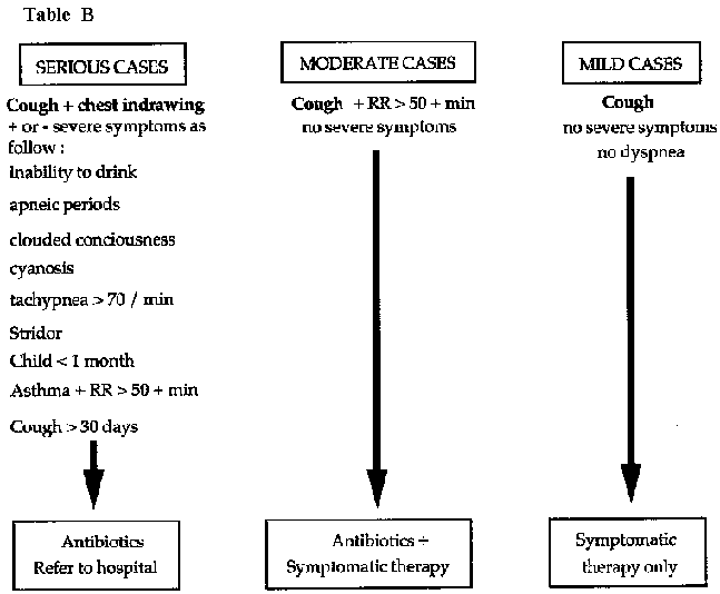
Table
Some upper respiratory tract infections require antibiotic treatment:
- Acute laryngitis: because of the severe dyspnea, this condition will be classified as a serious case and will receive antibiotics.
- Tonsillitis et otitis media: cough is often associated for antibiotic indication.
MEASURES FOR PRESENTING LOWER RESPIRATORY TRACT INFECTIONS IN THE UNDERFIVES
- Improve environment (better housing, less crowding).
- Bedding, blankets, clothing.
- Better nutrition.
- Immunization against measles, pertussis and diphtheria: Expanded Program of Immunization (EPI).
Common cold
Viral infection of the nasopharyngeal mucosa which are frequent and seasonal. Person to person transmission is usually airborne.
Clinical features
- Runny nose, often with fever and cough.
- May be the prodrome of influenza or measles.
- Sometimes accompanied by conjunctivitis.
Treatment (dispensary)
- Nasopharyngeal lavage using a syringe filled with normal saline (or clean water with ORSadded, 1 sachet/litre), 4 to 6 times a day.
- Treat fever.
- Treat or take preventive steps against conjunctivitis.
- If allergic component (morning sneezing fits):
promethazine
(PO)
Adult: 75 mg/d divided in 3 doses x 3-5 days
Child: 1 mg/kg/d divided in 3 doses x 3-5 days
or
chlorphenamine: 12 mg/d divided in 3 doses x 3-5 days
Follow-up
Risk of secondary infection and acute otitis media in infants. Always check the tympanic membranes of an infant with a cold.
Pharyngitis and Tonsillitis
Infection and inflammation of the pharynx and tonsils accompanied by fever, dysphagia and adenopathy.
Treatment
The two main objectives of therapy are to recognise and treat the tonsillitis of diphtheria and to reduce the complications of streptococcal throat infections (acute rheumatic fever and cardiac lesions).
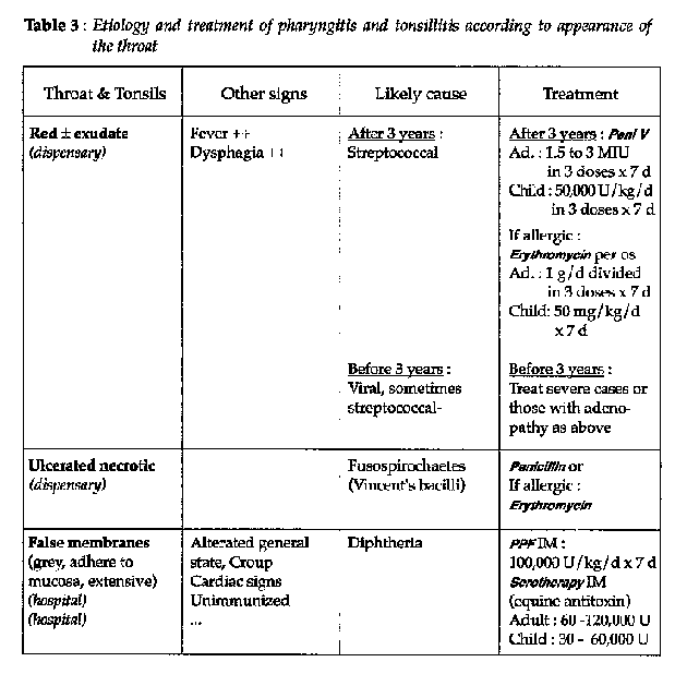
Table 3
- Always treat the fever and keep well hydrated (dysphagia).
- Patients with infectious mononucleosis will almost always present with an allergy to ampicillin. Stop the treatment.
- Follow-up to exclude acute rheumatic fever (polyarthritis, cardiac signs) and glomerulonephritis (edema, proteinuria, hypertension, hematuria).
- In case of diphteria, procede a survey in the patient neighbourhood. Contacts should be systematically treated with penicillin or erythromycin.
Note
Test the sensitivity to the equine antitoxin (SC 0.1 ml), wait 20 minutes to check an adverse reaction before complete treatment.
Acute otitis
Otitis externa
Infection of extemal auditory meatus (sometimes due to a foreign body).
Clinical features
- Pain, elicited especially by traction upon the pinna.
- Redness of meatus + abscess.
- May be an exudate.
- Drum normal.
Treatment (dispensary)
- Analgesia: acetylsalicylic acid or paracetamol.
- Local: if exudate ravage with normal saline. Apply gentian violet with a cotton bud for 3-5 days.
- If present, remove the foreign body.
Otitis media
Acute infection of the middle ear. Usually bacterial, tracking up from the nasopharynx: streptococcal, pneumococcal, Hemophilus influenzae in children under 5 years.
Clinical features
- Fever, severe pain, crying, agitation, vomiting, diarrhea.
- Ear drum: becomes progressively congested, inflamed, bulging, and finally perforates with release of pus.
Treatment (dispensary)
- Treat for fever and pain.
- If upper respiratory tract infection: nasopharyngeal ravage (ringer lactate).
- Rehydration if necessary.
-Antibiotherapy:
Over 5 years
|
Adult: |
penicillin v(PO): 2 MIU/d divided in 3 doses x 10 days |
|
Child: |
penicillin V(PO): 100,000 IU /kg/d divided in 3 doses x 10 days |
| |
or |
| |
PPF (ou procain penicillin): 100,000 IU/kg/d IM x 3 days, then peni V per |
|
|
os: same dose divided in 3 doses/day (total treatment: 10 days) |
If allergic to penicillin:
erithromicin (PO):
|
Adult: |
1.5-2 g/d divided in 3 doses x 10 days |
|
Child: |
50 mg/kg/d divided in 3 doses x 10 days |
Under 5 years
ampicillin (PO): 100 mg/kg/d divided in 3 doses x 10 days
or
cotrimoxazole (PO): 60 mg/kg/d SMX divided in 2 doses x 10 days
- Paracentesis: is indicated if the ear drum is bulging but not yet perforated. Should be done in the infero-posterior quadrant.
Aspirate the pus and prescribe an antibiotic as above.
Prognosis
If neglected, acute otitis media may become chronic. There is also a risk of mastoiditis.
Chronic otitis
Chronic infection of the middle ear with perforation of the tympanic membrane.
Clinical features
-Chronic discharge (otorrhea)
-Occasional acute re-infection: fever + pain, usually associated with an obstruction to drainage through the perforated drum with secondary infection by streptococci, pneumococci or gram negative organisms.
Treatment (dispensary)
-Do not prescribe antibiotics.
-Only if acute re-infection occurs give:
ampicllin
or
cotrimaxozole
in the same doses as for acute otitis media.
Lavage with
normal saline and aspirate with a syringe.
-Always put a little dry cotton wool or small wick in the ear to absorb the discharge; change 3-4 times/day til dried up.
Prognosis
-Risk of deafness in affected ear.
-Risk of mastoiditis and meningitis during acute re-infections.
Acute laryngitis
Acute infections of the laryngeal mucosa often associated with viral infections (e.g. colds, measles...).
Prognosis
The prognosis is good. However, patients sometimes develop partial respiratory obstruction, and it is important to identify these "high risk' situations and to take the necessary precautions.
ADULT
- Usually associated with a "hoarse" voice and a cold. The etiology is viral. Symptomatic treatment: A A.S. or paracetamol(PO).
- Rarely, epiglottitis from H. influenzae, diphtheria or retropharyngeal abscess. In these cases, use the same methods as for treating children.
- Tuberculous laryngitis.
CHILD
There is a risk of respiratory obstruction.
Signs of distress
Inspiratory stridor, with or without intercostal recession, pallor, with or without cyanosis with cough and "croupy" voice.
There are 2 distinct clinical features.
1. Progressive dyspnea (1 or more days)
In a child < 3 years, if other causes have been eliminated (e.g. diphtheria, retropharyngeal abscess, foreign body), the dyspnea is probably due to mild subglottic obstruction from a viral infection (laryngo-tracheobronchitis).
It is important to watch the child carefully, to keep him calm and to provide humidified air.
Antibiotics are unnecessary except for secondary infections (use PO ampicillin or cotrimoxazole). Steroids are not useful.
If the dyspnea worsens, intubation or tracheostomy may be necessary.
2. Rapid onset dyspnea (several hours)
Carefully examine the patient in a sitting position. Do not lie them down.
-Foreign body: if the dyspnea becomes labored, remove foreign body rapidly, in surgical surroundings.
-Acute epiglottitis from Haemophilus influenzae
· Child of 3 - 8 years: sudden onset dyspnea, high fever, stridor, dysphagia (drools saliva), breathes through mouth, cervical lymphadenopathy.
· Do not lie the patient down and avoid examining the larynx as these actions may precipitate respiratory obstruction.
· Keep the child sitting in a humid atmosphere.
Give:
ampicillin (IV): 200 mg/kg/24 hours divided in 3-4 injections,
reverting to oral treatment as soon as possible; total duration: 7
days
or
chloramphenicol(IV): 100 mg/kg/d divided in 3-4 injections,
reverting to oral treatment as soon as possible; total duration: 7 days
· Severe distress or obstruction: tracheostomy.
-Recurrent laryngitis
· Child of 2-4 years with a cold or measles.
·
Nocturnal dyspnea with no fever.
· Place the infant in humidified
atmosphere.
· Eventually, give: promethazine(PO): 75 mg/kg/d divided in
3 doses x 5 days
or
chlorphenamine (PO): 12 mg/d divided in 3 doses x 5
days
-Diphtheria: false membrane in the throat
· Unvaccinated children.
· Sometimes the false
membrane is extensive an adherent.
· Poor general condition.
·
Treatment:
diphtheria antitoxin
penicillin G or PPF IM
· Tracheostomy if necessary.
Sinusitis
Infection of the sinus mucosae with purulent nasal discharge. May originate from:
- the nose: rhinitis, allergic rhinitis, nasal obstruction (e.g. mal-formation, trauma);
- the teeth: caries with arthritis and /or osteitis.
Clinical features
Associated with pain and a purulent nasal discharge.
ADULT
- Pain
· Periorbital: frontal sinusitis.
· Facial:
maxillary or ethmoidal sinusitis.
- Purulent unilateral nasal discharge on the affected side with nasal obstruction and a moderate fever.
- Examination:
· Exquisite tenderness can be elicited over these
points.
· Rhinoscopy: inflamed mucosa with purulent exudate.
Bacteria responsible are Haemophilus influenzae in persons < 5 years and pneumococcus in older persons.
INFANTS
Acute ethmoiditis: high fever, edema of lower eyelids and the bridge of the nose with purulent rhinorrhea.
Danger of spread to bone or orbit. Treat vigorously.
Bacteria responsible are Haemophilus, pneumococcus and staphylococcus.
Treatment (dispensary)
- Nasopharyngeal lavage with removal of foreign body (if found).
- A.A.S. or paracetamol for fever and pain.
- If dental focus of infection, extract tooth under antibiotic cover.
- Antibiotic:
cotrimoxazole (PO): 60 mg of SMX/kg/d divided in 2 doses x 10 days
or
ampicillin (PO): 100 mg/kg/d divided in 3 doses x 10 days
- Ethmoiditis
ampicillin (IV): 200 mg/kg/d divided in 3 or 4 injections stat until cured. Change to PO as soon as possible.
or
chloramphenico/(IV or IM): 100 mg/kg/d divided in 3 or 4 injections, then change to PO as soon as possible.
Prognosis
Acute sinusitis may become chronic, so always exclude other pathology (e.g. foreign body, allergy, dental caries...).
Bronchitis
Acute bronchitis
Acute infection of the bronchial mucosa
Clinical features
- Often preceded by an upper respiratory tract infection.
- Cough, dry at first, then productive.
- Low grade fever.
- No marked dyspnea.
- Scattered rhonchi.
Treatmert (dispensary)
-In basically healthy patient following rhino-pharyngitis or flu.
· Keep well hydrated, treat fever, humidified air if possible.
· Nasopharyngeal lavage with isotonic solution(normal saline or ringer lactate).
· No antibiotics (mostly viral).
-In patient with poor basic health (malnutrition, measles, rickets, anaemia, chronic bronchitis, cardiac disease, elderly...) or dyspnea > 50 mn or other serious signs.
In these cases, superinfection is probable (haemophilus, gram -
bacilli, pneumococcus). Treat with:
cotrimoxazole (PO)
Adult: 1,600 mg/ d of SMX divided in 2 doses x 5-7 days
Child: 60 mg/kg/ d of SMX divided in 2 doses x 5-7 days
or
ampicillin (PO): 100 mg/kg/d divided in 3 doses x 5-7 days
or
chloramphenicol (PO): 50 mg/kg/d divided in 3 doses x 5-7 days
- Where wheezing occurs, treat as asthma.
Chroniques
Chronic inflammation of the bronchial mucosa of irritant (tobacco) or allergic (asthma) origin, progressing towards chronic respiratory failure.
Part of the syndrome of chronic obstructive airways disease (COAD).
Clinical features
- Morning cough, clear sputum, bronchial rales.
- If secondary infection: fever and purulent sputum.
- Always exclude TB: sputum smear for AFB.
Treatment (dispensary)
- Discourage cigarette smoking.
- No antibiotics unless secondary infection. In this case, see acute bronchitis.
Pneumonia and Bronchopneumonia
Infection of pulmonary alveoli and bronchial mucosa.
Cause:
- viral
- bacterial: pneumococcus, Haemophilus influenzae,
mycoplasma pneumonia
- parasitic: pneumocystis carinii (AIDS)
Clinical features
- High fever (> 39°), cough, respiratory distress, chest pain and tachypnea (> 50/min).
- Examination: dullness to percussion, diminished vesicular breath sounds, crepitations and sometimes bronchial breath sounds.
Treatment
Depends on age and presence of respiratory distress tachypnea (> 60/mn in infants less than 2 months, > 50/mn from 2 to 12 months, > 40/mn from 1 to 5 years), intercostal recession, alar flare, stridor, cyanosis, respiratory pauses, xyphi-sternal recession.
Other serious extrapulmonary signs can be present.
ABSENCE OF SERIOUS SIGNS
- Classical pneumonia in adults and children < 5 years
Localised crepitation, sometimes bronchial breathing or localised dullness to percussion = pneumococcus. By far the most common germ after 5 years of age.
Treatment (dispensary)
penicillin V(PO):
Adult: 2,4-3,6 MIU/d divided in 3 doses (tab 250 mg = 0.4 M1U: 2-3 tab x 3/d) x 5 days
Child: 50 000 IU/kg/divided in3 doses x 5 days
or
cotrimoxazole (PO):
Adult: 1600 mg of SMX/divided in2 doses x 5 days
Child: 50 mg of SMX/kg/divided in 2 doses x 5 days
- Pneumonia in child of 2 months to 5 years
H. Influenzae
common at this age. Therefore, first line of treatment:
cotrimaxazole(PO): 50 mg of SMX/kg/d divided in 2-3 doses x 5 days
or
ampicillin(PO):100 mg/kg/d divided in 3-4 doses x 5 days
or
amoxycillin (PO): 50 mg/kg/d divided in 3 doses x 5 days, depending on availability
- Pneumonia in infant < 2 months
Hospitalize (risk of rapid decompensation).
ampicillin PO if
possible (if not IM): 100 mg/kg/d divided in 3-4 doses x 7 days
Always treat fever and ensure adequate hydration and nourishment. Always review the patient 2 days later.
PNEUMONIA WITH RESPIRATORY DISTRESS: HOSPITALIZE
- Adult and child > 5 years
· If clinical evidence favours pneumococcus (one or several
systematic foci with crepitation and/or decreased vesicular breath sounds,
sometimes bronchial breathing or dullness to percussion):
PPF IM:
|
Adult: |
3-4 MIU/d in 1 injection x 2-3 days |
| |
then commence oral therapy with peni V: 3-4 MIU/d divided in 3-4 |
| |
doses to complete 7 days |
|
Child: |
50.000 UI/kg/d in 1 dose x 2-3 days |
| |
then commence oral therapy with peni V: 50.000 IU/kg/d divided in 3-4 |
|
|
doses to complete 7 days |
or
chloramphenicol IV-IM:
|
Adult: |
3-4 g/d divided in 3-4 doses over several days, then commence orally |
|
|
(same dosage) to complete 7 days |
|
Child: |
100 mg/kg/d divided in 3-4 doses over several days, then commence |
|
|
orally (same dosage) to complete 7 days |
· In all other cases:
chloramphenicol IV or IM:
|
Adult: |
3-4 g/d divided in 3-4 doses over 2-3 days |
|
Child: |
100 mg/kg/d divided in 3-4 doses over 2-3 days, then in both cases |
|
|
change to oral treatment with the same dosage to complete 7 days |
or
ampicillin IV or IM:
|
Adult: |
3-4 g/d divided in 3-4 doses over 2-3 days |
|
Child: |
100 mg/kg/d divided in 3-4 doses over 2-3 days, then in both cases |
|
|
change to oral treatment with the same dosage to complete 7 days |
Where no improvement with ampicillin after 2 days, combine
with
gentamicin IM:
|
Adult: |
160 mg/d divided in 2 doses |
|
Child: |
3-6 mg/kg/d divided in 2 doses x 7 days |
- Child of 2 months to 5 years
chloramphenicol IV or IM: 100 mg/kg/d divided in 3-4 doses; change to oral treatment as soon as possible in the same dosage to complete 7-10 days
or
ampicillin IV or IM: 100 mg/kg/d divided in 3-4 doses; change to oral treatment as soon as possible in the same dosage to complete 7-10 days
When possible, combine with gentamicin IM: 6 mg/kg/d divided in 2 doses during 7-10 days
In the absence of improvement or when deterioration occurs at the end of properly conducted treatment, think about staphylococcal pneumonia.
- Infant < 2 months
ampicillin IV or IM: 100 mg/kg/d
divided in 3-4 doses; change to oral treatment as soon as possible in the same
dosage to complete 7-10 days
plus gentamicin IM: 6 mg/kg/d divided in 2-3
doses x 7-10 days (for neonates < 10 days old: 4 mg/kg/d in 2 doses))
When no improvement occurs or there is deterioration after 4 days of correct treatment, think about a staphylococcal pneumonia (see "staphylococcal pneumonia").
In all cases, treat the temperature, ensure adequate nutrition and hydration (gastric tube if necessary). If oxygen available, use by means of nasal tube at the rate of 1 litre per minute when there is respiratory distress.
REFRACTORY PNEUMONIA IN ADULTS OR OLDER CHILDREN
Consider atypical pneumonia (mycoplasma) or tuberculosis. Alternative therapies to try: tetracycline
Adult: 1.5-2 g/ d divided in 3-4 doses x 7-10 days
Child > 8 years: 50 mg/ kg/ d divided in 3-4 doses x 7-10 days
or
erythromycin: same dosages as for tetracycline
or
cotrimoxazole
Adult: 1600 mg of SMX/d divided in 2 doses x 7-10 days
Child: 50 mg of SMX/kg/d divided in 2 doses x 7-10 days
If at the end of 3 courses of therapy the signs persist, consider tuberculosis (see "Tuberculosis").
Staphylococcal pneumonia
Staphylococcal pneumonia often occurs in an infant that is otherwise unwell (malnutrition, skin sepsis...).
Clinical features
- Fever, pallor, fatigue.
- Signs similar to those of severe bronchiolitis, with vomiting, diarrhea, abdominal distension, often skin abscesses.
- Auscultation: asymmetrical chest signs + pleural effusion.
- Neutrophilia.
- Chest X-ray: bullae, pleural effusion.
Treatment (hospital)
- Antibiotics, if available:
cloxacillin (IV): 100 mg/kg/ d divided in 4 injections x 10 days
and
gentamicin (IM): 3-6 mg/kg/d divided in 2 injections x 10 days
Otherwise:
chloramphenicol(IV): 100 mg/kg/d divided in 3 injections x 10 days
- Hydration: oral or IV.
- If there is a significant effusion, a pleural tap may be necessary or, if severe, an intercostal catheter with underwater drain.
Prognosis
Danger of complications of suppurative pleurisy, pneumothorax and pyo-pneumothorax.
In a pediatric ward where staphylococcal pneumonia are expected to be managed, health workers should be trained to perform urgent pleural tap. Adequat equipment should always be available.
Bronchiolitis
- Acute viral infection of the bronchioles occurring in infants under 10 months of age which can lead to fatal acute respiratory failure.
- Tends to occur in epidemics during the cold season.
Clinical features
- Onset often follows a cold.
- Low grade fever, cough, variable degree of respiratory distress with tachypnea, alar flare and chest indrawing (stemal and intercostal recession).
Cyanosis if severe.
- Hyperinflated chest, hyper-resonant to percussion.
- Auscultation can be normal or reveal rhonchi (wheezes) and crepitations.
Treatment (hospital)
- Close monitoring: very important.
- Sitting position (propped up or held by mother).
- Keep well hydrated but avoid fluid overload.
Humidified air.
- Bronchodilators: try salbutamol as a therapeutic test if available. The least dangerous is the spray (see "Asthma", see 67). Make two attempts at 15 minutes intervals, then wait. If there is improvement, continue; if not, do not persist.
- Corticosteroids not effective.
- Antibiotics to prevent secondary bacterial
infection:
cotrimaxazole (PO): 40 mg of SMX/kg/day in 2 divided doses x 5 days
or
ampicillin (PO or IM): 100 mg/kg/d divided in 3 doses x 5 days
- If cardiac decompensation (gallop rhythm, rate > 160): digoxin (IV): 0.01 mg/kg stat every 6-8 hours for the first 24 hours thence same dose once daily as maintenance. furosemide(IV):1 mg/kg
- Respiratory fatigue: if possible, intubation and manual ventilation.
Prognosis
- May have high mortality rate.
- Possibility of recurrence.
Asthma
Paroxysmal reversible airways obstruction due to a combination of bronchospasm, peribronchial edema and hypersecretion. Often allergic in origin.
Clinical features
- Wheeze (prolonged expiratory phase).
- Cough, dyspnea.
- Auscultation: expiratory rhonchi (wheezes) in both lung fields.
- 3 forms:
· simple attack,
· unstable asthma: repeated
attacks,
· status asthmaticus: prolonged, severe attack.
Treatment
Certain intestinal parasites during their invasive phase can cause allergic phenomena such as urticaria or asthma. Always think of this and treat in an endemic area: hookworm, strongyloides, ascaris, schistosomes, filaria.
SIMPLE ATTACK (dispensary)
- Half-sitting position, reassurance, hydration, oxygen if available.
- Aminophylline (PO): 5 mg/kg every 6 hours as
necessary
(contra-indicated in children under 2 years).
- Child < 2 years: commence with salbutamol if available (the spray is the least dangerous), if not adrenaline (epinephrine) (see "alternative treatments"). Aminophylline should only be used as a last recours at this age.
SEVERE CASES = STATUS ASTHMATICUS (hospital)
- Aminophylline (IV): 5 mg/kg diluted in 100 ml of 5% glucose, injected over 20 to 30 minutes, then 5 mg/kg over 6-8 hours depending on the clinical result.
- Never inject aminophylline undiluted (risk of convulsions and cardiac arrest).
- Combine with:
· Infusions (alternative glucose end ringer
lactate)
· salbutamol spray if avaiblable.
dexamethasone (IV):
Adult: 16-24 mg/d divided in 4-6 injections
Child: 0.1-0.5 mg/kg/d divided in 4-6 injections
Adjust the dosage to clinical state and decrease progressively.
- Antibiotics:
Adult: peni V PO or PPF IM: 3-4 MIU/d x 5 days
Child < 5 years: chloramphenicol IVor PO: 100 mg/kg/d divided in 3-4 doses x 5 days
or
ampicillin IV or PO: 100 mg/kg/d divided in 3-4 doses x 5 days
- After 2-3 hours aminophyllin infusion, when no improvement, return if possible to salbutamol or adrenaline.
UNSTABLE CASES
- Attacks which stop and recur despite treatment.
- Institute the following
aminophylline (PO)
Adult and child: 8-12 mg/kg/d divided in 3 doses, reduce dose progressively over several days
Duration depends on clinical state; decrease must be very gradual every 4-5 days.
Or better, but rarely available:
salbutamol(PO)
Adult: 12-16 mg/d divided in 3-4 doses x 5 days
Child: 0.2 mg/kg/d divided in 3-4 doses x 5 days, then decrease very gradually (every 4-5 days)
- In case of failure, consider short term, corticosteroid
therapy.
If used, it is essential to exclude underlying pulmonary
infection
(if in doubt: peni Vor cotrimoxazole).
Give prednisone (or
prednisolone) per os
Adult: 30 mg/d in a single dose x 3-4 days
Child: 1
mg/kg/d in a single dose x 3-4 days.
Then decrease very gradually every 3-4
days depending on the clinical state.
ALTERNATIVE TREATMENTS
- salbutamol spray: a double puff if an attack occurs, without exceeding 3-4 doses per day.
- salbutamol IV: difficult to control (tachycardia +++). Begin with 0.1 mg/10 kg in 100 ml 5 % glucose, over 20 minutes. Watch pulse, blood pressure. Continue with 1 mg/hour (0.25-1.5 mg/h) for adult; in child, 0.3-0.6 mg/10 kg/ hour. Change to oral treatment after 24 hours. Reserve for refractory cases in children or status asthmaticus.
- epinephrine (adrenaline) SC: use 0.1 % (1 mg/ml) solution.
Adult: 0.5 ml SC, repeat if necessary 30 minutes later; do not exceed 4 injectionsper day.
Child: 0.01 ml/kg SC without exceeding 0.5 ml/injection, repeat after 30 minutes if still necessary; do not exceed 4 injections per day; exercise extreme caution (+++) in infants less than 1 year old.
Watch pulse, blood pressure (tachycardia +++).
Adrenaline can be utilised in case of simple attack.
Tuberculosis
Disease of variable manifestations caused by Mycobacterium tuberculosis. It is important to understand the distinction between:
- Tuberculous infection: presence of M. tuberculosis in the organism, manifested by a positive skin test. Is very often asymptomatic.
- Tuberculous disease: affects about 10% of the infected population. Clinical disease can take very diverse forms:
· meningitis,
· pulmonary TB: the commonest form and the main source of transmission ("open" cases coughing up large numbers of AFB),
· lymphadenitis,
· osteo-articular, Pott's disease,
· intestinal, renal, peritoneal, cutaneous
- Transmission and maintenance of endemicity depend upon:
· the number and sources of infection: open pulmonary cases
can be easily identified by 3 consecutive daily sputum examinations
(direct
smear for AFB);
· living conditions: crowding, hygiene,
· susceptibility of the population (e.g. malnutrition).
- Individuals with low immune defences (e.g. malnourished, infants, elderly, AIDS patients) are more susceptible to the very severe forms (TB meningitis, miliary TB).
Note: not all cases of hemoptysis are necessarily TB. It is important not to forget other causes, especially if sputum smears are negative: paragonimiasis and meliodosis in Southeast Asia; systemic mycoses; histoplasmosis; and bronchogenic carcinoma.
- An active pulmonary TB is considered as an opportunistic among
AIDS or HIV infected patients. thus TB can be the initial step of
AIDS.
Control of tuberculosis
ENVIRONMENT
Improvement of living conditions in a community lessens the risk of contagion (e.g. ventilation, light, no crowding...).
BCG AND CHEMOTHERAPY
- The BCG vaccination confers limited individual protection, and is mainly effective against infantile TB meningitis. It does not protect against most other forms of TB and does not confer herd immunity upon the population.
- The chemotherapy of TB is a complex issue. Certain fundamental rules must always be followed:
- Chemotherapy is only effective if rigorously organized and controlled. On an epidemiological level, a poorly organized program may be worse than no program at all. Poor treatment compliance leads to chronic, refractory cases and encourages the emergence of drug resistance.
- The success of a program depends less upon the intrinsic quality of the chosen treatment regimen than upon rigorous supervision and follow-up of patients during the entire duration of therapy.
· The program should be designed specifically for the local social, cultural and economic conditions.
· TB control is almost always co-ordinated at a national
level.
Foreign medical beams must adhere strictly to the national guidelines.
Practical organization
- The objective is to reduce transmission. The way to achieve this is to find and to treat sputum-positive cases.
- Clues to the identification of infectious patients are:
· cough of more than 3 weeks duration,
·
hemoptysis, chest pain,
· weight loss,
· night sweats?
- Chest X-ray is not a pafficularly useful criterion for deciding upon treatment; only sputum-positive and extrapulmonary cases should be commenced on therapy.
- The laboratory must be capable of carrying out direct smears
for
AFB, otherwise a control program cannot be envisaged. Active case
detection is important, but only if a mechanism for effective treatment and
follow-up exists.
- The main issue is to establish the mechanisms (infrastructure, trained personnel, transport, supplies) that will ensure good treatment compliance and follow-up, whatever the particular regimen. Without this, the program will fail. The availability of sophisticated drugs (such as rifampicin) is an issue of much lesser importance.
Three examples of treatment regimens
Low COST
Cost of full course < $US 20; 12 months duration.
- isoniazid(5mg/kg/d) + thioacetazone(2.5mg/kg/d) combination= INH+ TB1
Adult: tab 300 mg INH + tab 150 mg TB1
Child (< 6 years): tab 100 mg INH + tab 50 mg TB1 each day by mouth for 12 months with an initial supplement of 2 months of streptomycin (IM): 20 mg/kg
HIGH COST
Cost of full course > $US 175; 6 months duration.
- isoniazid:
Adult: 5mgtkg/d
Child: 10 to 20 mg/kg/d
+ rifampicin: 10 mg/kg/d
+ pyrazinamide: 25 mg/kg/d for first 2 months only
+ ethambutol: 20 mg/kg/d for first 2 months only.
INTERMEDIATE COST
Cost of full course approx. $US 85; 8 months duration.
- First 2 months: isonazid: 5 mg/kg/d
+ rifampicin: 10
mg/kg/d
+ pyrazinamide: 25mg/kg/d
+ streptomycin(IM): 20 mg/kg/d
- Next 6 months: isoniazid + thioacetazone: 5 mg/kg/d of INH.
Note
- Consult other documents on tuberculosis control programs,
especially concerning case detection and short-course regimens
(40).
- Hemoptysis is not always caused by tuberculosis. Other causes
if
AFB -; paragonimosis and melioidosis in South East Asia; deep mycosis:
histoplasmosis; bronchopulmonary
cancer.
|
| |||||||||||||||||||||||||
Clinical Guidelines and Treatment Manual (MSF, 1993, 319 p.)
Chapter 3 - Gastro-intestinal diseases
Stomatitis
Inflammation of the oral mucosa, with or without infection, frequently found in infants.
If severe can contribute to malnutrition. Always treat carefully, and explain treatment to the mother.
Clinical features
- Sore mouth, dysphagia, anorexia, nausea, vomiting.
- Depending on etiology: red mucosa, aphthous or other ulcers, vesicles, white plaques.
Etiology and treatment
CANDIDA ALBICANS ("THRUSH") (dispensary)
- Common in infants.
- White plaques.
- Clean the mouth with a gauze swab soaked in sodium bicarbonate, then apply gentian violet with a cotton bud. Show the mother how to do this and have her repeat it 6 times a day.
- Often associated with gastro-intestinal candidosis. Treat all cases of oral thrush with: nyststin(PO): 100,000 to 200,000 IU/d divided in 3 doses x 5 to 10 days.
(Use the vaginal tablets if only these are available.)
- Educate the mother about oral hygiene.
- In severe forms, think of HIV infection.
HERPES SIMPLEX (dispensary)
Commoner in older children and adults. Infection causes pain and difficulty eating. Transmission is via microdroplets of saliva. Attacks are often precipitated by a febrile illness or stress.
- Oral toilet and apply gentian violet.
- Continued feeding and ensure good hydration.
- Treat any underlying illness (e.g. malaria, pneumonia).
With a secondary infection (rare if good oral
toilet):
cotrimoxazole (PO)
Adult: 1,600 mg/d of SMX divided in 2 doses x 5 days
Child: 40 mg/kg/d of SMX divided in 2 doses x 5 days
or
chloramphenicol(PO): 50 mg/kg/d divided in 3 doses x 5 days
SCURVY (dispensary)
Hemorrhagic stomatitis with bone and joint pains in the lower limbs (due to subperiosteal hemorrages). Caused by dietary vitamin C deficiency.
Local treatment: oral toilet and gentian violet
Curative treatment
ascorbic acid (vitamine
C)
Adult: 500-1000 mg/d divided in 3 doses during 2 to 3 weeks
Child: 100-300 mg/d divided in 3 doses during 2 to 3 weeks
Preventive treatment
ascorbic acid (vitamine
C)
Adult: 100mg/d
Child: 30-50mg/d
Nutritional education and supplementation with fresh fruit.
Other causes
Vincent's angina
Measles (Koplik's spot)
Diphtheria
Scarlet fever (strawberry tongue): a streptococcal
infection.
Treatment:
PPF(or procain penicillin): 100,000 IU/mg/d in a single
injection x 5 days
then
penicillin V(PO): same dose divided in 3 doses/d x
10 days
Angular stomatitis of the lips: deficiencies in iron and various vitamins:
multivitamins
and/or
ferrous sulphate +folic
acid.
Gastritis and Peptic Ulcer
Inflammatory or ulcerative lesions of the gastro-duodenal mucosae.
Clinical features
- Epigastric burning pain, sometimes made worse and sometimes relieved by food (especially milk) but recurring about two hours after meals.
- Acid regurgitation, nausea.
- Abdomen soft and non-tender (unless perforation).
- Exclude parasitosis (strongyloides): stool examination.
Treatmert (dispensary)
- Diet: avoid spices, alcohol, tobacco, carbonated
drinks.
Encourage regular meals, dairy products.
- Antacids: aluminium hidroxide (PO): 300 to 500 mg in a single dose, taken 1 hour after each meal or during attacks of pain
- Reassure the patient: anxiety may be a causative factor. If needed: diazepam (PO): 15 mg/d divided in 3 doses for a brief period (5-10 days)
- If severe pain continues, exclude perforation: examine abdomen for peritonism, PR exam for rectal blood (melena on glove), keep under observation, surgical referral if necessary.
Give: atropine (IM or SC): 1 mg stat.
(hospital)
- If hemorrhage:
· establish IV line,
· give plasma volume expander
(Haemacel...),
· nasogastric tube: to observe if hemorrage
continues,
· transfuse if possible and refer to a surgical unit.
NB: acetylsalicylic acid and other non-steroidal anti-inflammatories are contraindicated in patients with a history of peptic ulcer.
Acute diarrhoea
Loose, frequent stools. Different cultures have different definitions, but as a guide, diarrhea means at least 3 loose or watery stools in a day.
Major complications:
- Dehydration: the principal reason for the mortality attributable to diarrhea
- Negative effect on nutritional status
Clinical assessment of the patient
HISTORY
- Duration of illness.
- Frequency and consistency of stools (blood, mucus).
- Frequency and duration of vomiting.
- Output, colour and quantity of urine.
- Fever or convulsions.
- Type and quantity of fluids and food ingested.
- Presence of blood or mucus in the stool.
- Presence of other cases in the household.
PHYSICAL EXAMINATION
- Temperature (rectal if possible).
- Respiration (acidosis: Kussmaul breathing).
- Weight:
· as a baseline to monitor rehydration,
· as an
indicator of degree of dehydration.
- Nutritional status.
CLINICAL EVALUATION OF DEGREE OF DEHYDRATION
See table 4.
STOOL EXAMINATION
Direct smear, if available, to look for trophozoites of entamaeba hystolitica or giardia lamblia.
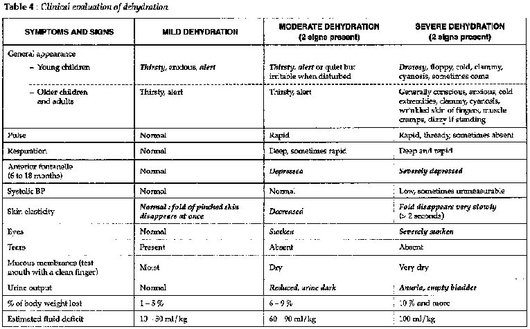
Table 4
Etiology

Table 5
Treatment
Basic principles:
- Prevent dehydration.
- Replace fluid if dehydration already exists.
- Maintain nutrition.
PREVENTION OF DEHYDRATION
Cases of diarrhea with no signs of dehydration:
- Advise increasing fluid intake (water, soup, juices, rice water).
- Encourage the use of home-made sugar / salt solutions.
- Continue breast feeding and normal diet.
- Warn mother to bring child back if:
· signs of dehydration appear (explain),
· diarrhea
persists.
FLUID REPLACEMENT
- Two tasks:
· Rehydration: correct the deficit in water and
electrolytes.
· Maintenance: replace continuing losses (diarrhea and
vomiting).
- Two methods of fluid replacement:
· ORS: for mild to moderate dehydration, give by mouth or by nasogastric tube if child unable or unwilling to drink.
· Ringer's lactate: for severe dehydration or if there is intractable vomiting.
- Quantities of fluid are calculated according to the condition of the patient (see tables 6 and 7). As a general rule, for severe dehydration 200 ml/kg/day should be given with the first half during the first 4 hours. For moderate dehydration, give 100 ml/kg/day with first half given during first 4 hours.
- Mild cases can be treated as outpatients, after the mother has been shown how to use ORS. Moderate and severe cases require supervision as to the evolution of the diarrhea and progress of rehydration.
- If it is impossible to place an IV line in a severely dehydrated child, fluids are sometimes given intraperitoneally or subcutaneously. However these techniques should not be encouraged, as they are less safe and no more effective than giving ORS by nasogastric tube.
Note: solution of salt-sugar: 2 pinches of salt (3 g), 4 tablespoons of sugar, or 8 pieces (40 g), dissolved in 1 liter of boiled water, cooled and with added fruit juice.
Table 6: Rehydration protocol
- The volumes indicated are guides only.
- Before using this table, consider all of the following:
· Rehydration must be evaluated in terms of clinical signs, not in terms of volume of fluids given.
· If necessary, the volumes given below can be increased or else the initial high rate of administrahon can be maintained until there is clinical improvement.
· Periorbital edema is a sign of fluid overload in infants.
· Maintenance therapy (table 7) should begin as soon as signs of dehydration have resolved, but not before.
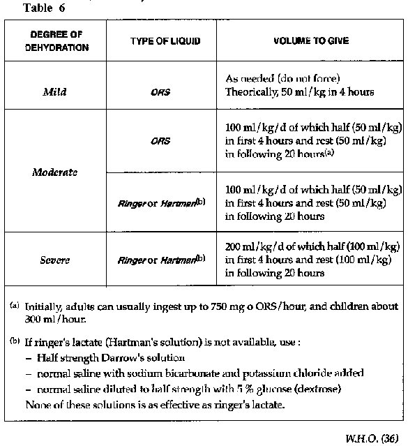
Table 6
Table 7: Maintenance therapy
Notes:
- fluids to be given after correction of dehydration;
- adapt re-hydration treatment to the clinical status of the patient;
- to avoid hyrpernatremia altemate ORS and water.
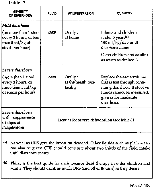
Table 7
MAINTAIN NUTRITION
It has been shown that there is no physiological reason for discontinuing food during bouts of diarrhea and that continued nutrition is beneficial to both adults and children. Continued feeding should be encouraged.
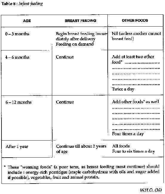
Table 8
MEDICINES
- Remember that 50-60 % of acute gastro-enteritis is viral (see table 5).
- Certain antibiotics are used to treat specific intestinal infections.
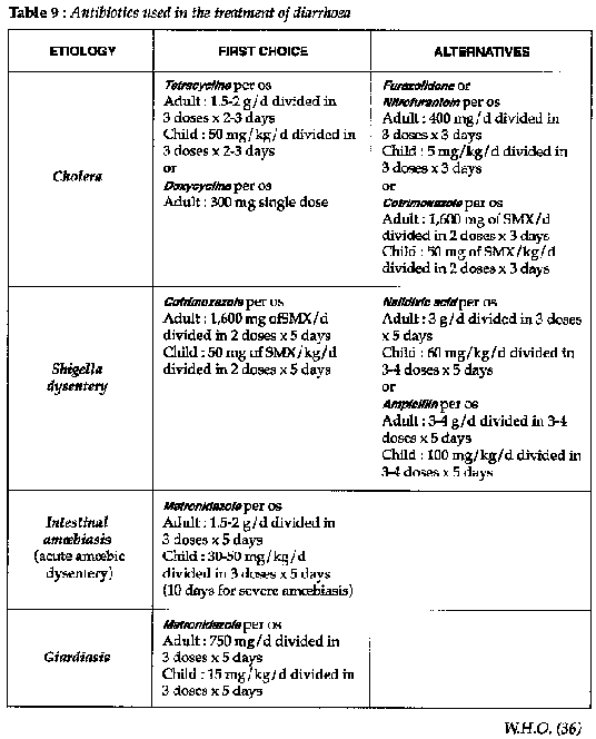
Table 9
- Other anti-diarrhoea indications (e.g. absorbents) are contraindicated in children.
- Always treat the fever and consider other causes for the diarrhoea (e.g. malaria, otitis, pneumonia).
Prevention of diarrhea
HEALTH EDUCATION
Directed at mothers in dispensaries, MCH clinics and feeding centers, at the time ORS is prescribed.
Take-home messages:
1) Breastfeeding:
· on its own up to age 4 months
· continue up to
age 2 years
2) Solid foods ("weaning foods" is a very poor term): introduce these from about age 4 months
3) Food preparation
4) Drinking water
5) Hygiene
SANITATION
- Provision of safe drinking water in sufficient quantities
- Disposal of feces
MASS CHEMOPROPHYLAXIS
- This is only ever considered in cholera epidemics. It is of doubtful efficacy in controlling an outbreak and can only be justified in a sequestered populations: a ship, a medium size where the attack rate is high (more than 2 %) and where it is possible to administer an effective prophylactic dose under supervision to the whole group concerned.
- In endemic situation, it can be given to close family contacts.
- Doxycyclineshould be choosen.
Note
Composition of ORS sachets (to be dissolved in 1 litre of clean water (do not tell mothers to boil the water as this is very expensive in terms of time and fuel, and also unnecessary). (See table C).
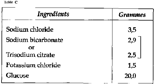
Table
|
| ||||||||||||||||||||||||||||||||||||||||||||||||||||||||||||
Clinical Guidelines and Treatment Manual (MSF, 1993, 319 p.)
Chapter 4 - Skin conditions
Dermatology
Infections and infestations are by far the most frequent forms of skin pathology in tropical countries. As well as treating affected individuals, it is important to consider these conditions as indicators of the general standard of hygiene and sanitation and to define appropriate public health interventions (provision of water that is adequate in quality and quantity, health education, soap...).
Clinical assessment of the patient
The physical examination:
- Describe the basic lesions:
· macules
· papules
· vesicles
·
bullae
· abscess
· pustules
· squames
· weeping
lesions
· crusts...
- Look for pruritis.
- Look for regional or systemic manifestations: lymphangitis, adenopathy, fever, septicemia, metastatic infection...
- Look for a cause: mosquito bite, jewelry, allergy, scabies, lice, otitis media.
- Consider the nutritional status and the general health of the family, particularly for infectious dermatosis.
Patients with dermatological conditions often present late. At
this stage, initial and specific signs are often replaced by infection. In these
cases, treating the overlying infection is not enough.
Patients should be
re-examined after the treatment of
infection.
Impetigo and other purulent dermatoses
Highly contagious skin infection (streptococcal or staphylococcal affecting mainly children of school age).
Clinical features
-Initially lesions located around orifices.
- Multiple crusty lesions, sometimes associated with pustules (one to several). Lesions are extended by scratching
- Acute impetigo produces bullous lesions.
- Streptococcal lesions are superficial, staphylococcal lesions are deep.
Treatment (dispensary)
- Cut fingernails, instruct mother to wash child daily with soap.
- Clean lesions with a disinfectant (chloramine or chlorhexidine-cetrimide solution; preparation: see table 25). Remove crusts, incise any abscesses.
- Apply gentian violet solution twice daily.
- On the scalp look for head lice or ringworm (tinea capitis).
- Explain treatment to the mother and treat other members of the family as necessary.
- Do not give antibiotics unless there are signs of regional or systemic spread. If so:
penicillin V(PO)
Adult: 2.4 MIU/d divided in 3 doses x 5 days
Child: 100,000 mg/kg/d divided in 3 doses x 5 days
If no improvement or extensive abscesses, staphylococcal
infection is likely. If available give: erythromycin (PO): 50 mg/kg/d divided in
3 doses x 5 days
or
cloxacillin(PO): 100 mg/mg/d divided in 3 doses x 5-7
days
- Carbuncles on the face. There is a danger of intracerebral
metastasis, so treat vigorously for 5-7 days.
If
available:
cloxacillin (IV or PO): 100 mg/kg/d divided in 4 injections x 7 days
or
chloramphenicol(IV): 75 mg/kg/d divided in 4 injections x 7 days
or
ampicillin (IV): 100 mg/kg/d divided in 4 injections x 7 days
+gentamicin (IM): 3 mg/kg/d divided in 2 or 3 injections x 7 days
Herpes simplex, Herpes zoster
Herbes simplex
Relapsing vesicular eruption due to Herpes simplex virus affecting the mucus membranes and skin. Facial infections may be serious if they involve the eye.
Treatment (dispensary)
- Clean the lesions with an antiseptic such as chlorhexidine-cetrimide solution (dilution: see table 25).
- If affecting buccal mucosa then treat as for stomatitis.
- If affecting the face encourage ocular hygiene.
- If generalized bacterial super infection, treat as for staphylococcal impetigo.
Herpes zoster
Acute dermatosis due to resurgence of the varicella-zoster virus, which also causes chickenpox. Preceded by severe neuralgic pain, the eruption is vesicular on an erythematous base and is almost invariably unilateral, occupying the dermatome of a peripheral nerve.
Treatment (dispensary)
As above, plus analgesia.
Scabies
Contagious skin infestation caused by a mite, Sarcoptes scabiei. Its occurrence is closely related to a lack of water and poor hygiene.
Clinical picture
- Nocturnal itching, scratch marks, burrows between the
fingers
(made visible by applying ink, then washing it off), and papules.
- Secondary infection (from scratching) resembles impetigo.
- Whole family is often infested.
- Often localised to: genital region, axillae, chest, breasts, hands and thighs.
Treatment (dispensary)
- Wash the whole body with a mild soap and dry, then apply 25 % benzyl benzoate emulsion (BBE) to the whole body except head and neck. Use a broad paint brush if available. Allow to dry, then put on the same clothes.
- Repeat for 3 days.
- If a secondary bacterial infection occurs, treat as for impetigo for 4 to 5 days. Only apply benzyl benzoate emulsion once all lesions are closed (is very irritant).
- Treat the whole family. After the treatment, boil and air all clothes and bedding.
- Warn patients that itching may persist for several weeks. This represents an allergic reaction to the dead mites, not treatment failure.
Leg ulcer
Erosive lesion of the skin, usually occurring on the lower leg caused by:
- vascular (venous and/or arterial) insufficiency,
- bacterial or parasitic infection,
- underlying metabolic disorders.
Phadegenic ulcers have no apparent cause, they extend and become chronic.
Treatment (dispensary)
- Clean with chlorhexidine-cetrimide or chloramin solutions (dilution: see table 25).
- Excise necrotic edges.
- Daily dressing.
- Rest with leg elevated.
- Give oral antibiotics if local treatment fails:
penicillin
V(PO):
Adult: 2.4 MIU/d divided in 3 doses x 5 days
Child: 100,000 IU/kg/d divided in 3 doses x 5 days
If no improvement, give:
erythromicin (PO): 50 mg/kg/d
divided in 3 doses x 7 days
- Skin graft if ulcer is large. Only graft after local treatment has rendered it clean and flat with red granulation tissue in the base.
Note
- Think of Guinea worm in endemic zones.
- Give tetanus toxoid.
Fungal infection
Infant's thrush
Clinical features
Erythema of the buttocks and perineum, sometimes "weeping". Caused by infection with Candida albicans.
Treatment (dispensary)
- Clean with usual soap or an antiseptic (chloramine or chlorhexidine-cetrimide; preparation: see table 25).
- Apply gentian violet solution twice daily.
- Avoid damp clothing (leave buttocks bare).
- Intestinal thrush often co-exists:
Nystatin (PO): 4-600,000 IU/d divided in 3 doses x 10 days (vaginal tabs might be used for this purpose)
Dermatoses
Highly contagious fungal infections. Prevalence is associated with the level of personal hygiene.
Clinical features
BODY
There are several different forms:
- "Ringworm"
- Pityriasis versicolor with depigmented patches.
- Erythematous lesions in the skin folds (e.g. axillae and groin).
HEAD
Associated with loss of hair. Highly contagious in families.
Treatment
BODY (dispensary)
- Wash, dry and then apply whitfield's ointment twice a day.
- Use griseofulvin only in extensive cases (see below).
HEAD (dispensary)
- Cut hair and then shave the head.
- Wash, dry and then apply gentian violet twice a day for several weeks.
- If treatment fails use:
griseofulvin (PO)
Adult: 500 mg to 1 g/d divided in 3 doses
Child: 10 mg/kg/d divided in 3 doses
Treat for 10 days (sometimes treatment has to be continued for 1 month).
- Examine all the family.
- A short course of griseofulvin may be effective for adults:
griseofulvin: 1.5 g taken at the same time as a greasy meal.
However, there is a risk of digestive problems and vertigo.
Anthrax
A bacterial zoonosis of herbivorous mammals that is transmitted to humans by skin contact with carcasses or animal products, and rarely by ingestion of undercooked infected meat.
Clinical features
- Pustule that develops into a black eschar surrounded by vesicles and an inflamed area, with regional adenopathy. Painless.
- May cause fatal septicemia.
- Intestinal and pulmonary forms exist.
Treatment (hospital)
penicillin: PPF(or procain penicilline) IM
Adult: 4 MU / d in a single injection x 5 days
Child: 100,000 U/kg/d in a single injection x 5 days
or
penicillin V
Adult: 4 MU/d divided in 3 doses x 7 days
Child: 100,000 U/kg/d divided in 3 doses x 7 days
or
chloramphenicol per os
Adult: 1.5-2 g/ d divided in 3 doses x 7 days
Child: 75 mg/kg/d divided in 3 doses x 7 days
or
tetracycline per os
Adult: 1.5-2 g/d divided in 3-4 doses x 7 days
Child > 8 years: 50 mg/kg/d divided in 3-4 doses x 7 days
Prevention
Look for the source of contamination and take measures to prevent further transmission.
Other skin conditions
Eczema (dermatitis)
- Erythema with crusting, scaling, itching, and desquamation.
- Look for a cause (e.g. irritants, allergic, fungal, family history).
Treatment (dispensary)
- Apply gentian violet solution. If infected, treat as for impetigo.
- If chronic, consider an eczematized scabies infestation and treat appro-priately.
Urticaria
- Rapidly developing itchy papules.
- Look for a cause (e.g. insect bites, drug allergy, invasive stage of a parasitic infection of ascaris, hookworm, strongyloides, schisto-somiasis or loa-loa).
Treatment (dispensary)
- Intense itching:
promethazine (PO): 75 mg/d divided in 3 doses x 5
days
or
chlorphenamine (PO): 12 mg/d divided in 3 doses
- Angiedema (laryngeal or pharyngeal
involvement).
dexamethasone (IV): 4 mg (repeat if necessary)
- If anaphylactic shock, see "Shock".
Kwashiorkor
- Multiple lesions: desquamation, bullas...
- To prevent secondary infection, clean lesions with either chloramine or chlorhexidine-cetrimide solution and apply gentian violet (see table 25).
Pellagra
Dermatosis affecting sun-exposed skin due to a dietary deficiency in niacin and/or tryptophane. Commonest in populations whose staple carbohydrate is maize.
Clinical features
- Classically, "three D's": dermatitis, diarrhea and dementia.
- The dermatitis is painful, more marked in sun-exposed areas of forehead, neck, forearms and legs... symmetrical.
- Diarrhoea and neurological symptoms indicating serious
illness.
Treatment (dipensary)
Use:
nicotamide (vitomin PP)
Adult: 300 mg/d
Child: 300 mg/d until healing is complete and in conjunction with a rich protein diet.
If unavailable, use:
multivitamins (PO)
Adult: 6 caps/d divided in 3 doses x 15 days
Child: 3 caps/d divided in 3 doses x 15 days
Provide nutritional education (e.g. diversify staples, advise vegetables such as beans and lentils).
Note
Leprosy may in some instances resemble a chronic dermatosis such as eczema or pityriasis versicolor. Whenever there is the slightest doubt, perform a full examination of the peripheral nerves and take specimens for microscopy.
Endemic syphilis, Yaws & Pinta
Non-venereal treponematoses, affecting bone, skin or mucosa and spread by direct contact. Their occurrence is associated with over crowding and poor hygiene.

Table 10
Treatment (dispensary)
- benzathine penicillin (IM)
Adult: 1.2 MIU in a single injection
Child: 600,000 IU in a single injection
- Note that syphilis (both primary and secondary) must be treated with double this dose of benzathine penicillin: 2.4 MIU IM stat.
- If allergic to penicillin: tetracycline or erythromycin (PO): 2 g/d or 50 mg/kg/d divided in 3 doses x 5 days (14 days for syphilis)
Prophylaxis
For all household contacts:
benzathine penicillin (IM)
Adult: 600,000 IU in a single injection
Child: 300,000 IU in a single injection
Leprosy
A chronic infectious disease caused by the Hansen bacillus
(Mycobacterium leprae) and affecting the skin, mucosae and peripheral
nerves.
Humans are the only significant reservoir of infection.
Clinical features
COMPLETE CLINICAL EXAMINATION
- Patient must be undressed.
- The entire skin surface must be examined.
- Note the appearance of any lesions.
- Test the sensation (fine touch and pin-prick) of the lesions.
- Palpate the main peripheral nerves to detect any hypertrophy.
- Examine peripheral nervous function: motor, sensory and proprioception.
- Examine the nasal mucosa to detect any chronic rhinitis.
MICROBIOLOGICAL EXAMINATION
Ziehl-Nielsen stain
- Scraped incision method to obtain tissue juice but no
blood.
Pinch a fold of skin with a Kocher forceps so as to make it bloodless,
incise and saape the scalpel blade along the inside of the incision.
· one specimen from the edge of a lesion.
· one
from the earlobe.
- Also take a nasal swab.
CLASSIFICATION (RIDLEY-JOPLING SYSTEM) (See table D)
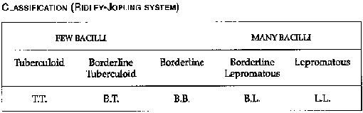
Table
Principles of management
The increasing frequency of strains of M. leprae resistant to dapsone poses a serious threat to leprosy control programs. The strategy of multiple drug therapy must be instituted in order to combat this problem.
It is patients with multibacillary forms of leprosy (BL and LL) who are most exposed to the risk of drug resistance and who are also most infectious to their household contacts.
The treatment of lepromatous leprosy has three objectives:
1. to reduce transmission
2. to cure the patient
3. to prevent the emergence of resistant strains of M. leprae
Patients with tuberculoid leprosy are less infectious but are more likely to suffer paralysis of peripheral nerves. The main objective of their treatment is to preserve function.
Patients under therapy are exposed to the risk of developing severe reactions. For this reason, as well as to ensure compliance, supervision is necessary. The program must be well planned, organized and adequately staffed.
Treatment
"Partially supervised" regimens work best. Daily doses of dapsone can be taken at home by the patient, but monthly doses of rifampicin should be administered by a health worker. The worker must ensure that the patient swallows the medication.
LEPROMATOUS LEPROSY (dispensary)
rifampicin (PO): 600 mg once per month, supervised
dapsone
(PO): 100 mg daily, at home
clofazimine (PO): 300 mg once per month,
supervised and 50 mg daily, at home
Duration of therapy: 2 years or more, depending upon progress
TUBERCULOID LEPROSY (dispensary - hospital)
rifampicin (PO):
600 mg once per month, during 6 months
dapsone (PO): 100 mg daily during 6
months (1-2 mg/kg/day)
- rifampicin must always be taken under supervision.
- dapsone may be taken at home. If treatment is interrupted, a full 6 months regimen should be completed after the medication has been discontinued.
REACTIONS
- Either reversal reactions (upgrading: shift towards TT end of spectrum) or erythema nodosum leprosum (ENL).
- Treat with clofazimine(PO): 100 to 300 mg/d
- If severe, use corticosteroids:
prednisone (or
prednisolone) PO: 80 mg D1; 75 mg D2; 70 mg D3; 65 mg D4; 60 mg D5...then
decrease 5 mg every
day.
|
| ||||||||||||||||||||||||||||||||||||||||
Clinical Guidelines and Treatment Manual (MSF, 1993, 319 p.)
Chapter 5 - Eye conditions
Conjunctivitis
- Acute inflammation of the conjunctivae which may be infectious (viral or bacterial), allergic or irritative.
- Infectious conjunctivitis is often endemic and may become epidemic in conditions of poor hygiene. Secondary infection may lead to keratitis and subsequent blindness.
- Viral conjunctivitis is often preceded by a cold.
Clinical features
- "Red eye" (injected conjunctivae), either unilateral or bilateral. May be purulent discharge. Visual acuity intact.
- Pain and photophobia are signs of comeal involvement. Look for pericorneal injection and examine after fluoroscein staining if available. Examine carefully to exclude foreign body (corneal or conjunctival).
- Chronic pruritis is usually the allergic form.
Treatment
(dispensary)
- Usual picture
· Wash a several times a day to remove any discharge. Use cooled boiled water or normal saline.
· Then, apply:
tetracycline eye ointment 1 %: 4 times/day x 5 days
or
sulphacetamide 10 %.
· Always look for foreign bodies (sub-conjuctival or comeal) and remove.
· Never use topical steroids.
(hospital)
- Ophthalmia neonatorum (gonococcal)
It is bilateral and appears immediately after birth. If only after 3 days, it is likely to be chlamydia.
· Prevention
Formerly, a 1% solution of silver nitrate was used for all neonates. This product is effective but may be dangerous if poorly prepared or stored; evaporation in hot climates may greatly increase the solution's concentration and thus toxicity. The current WHO recommendation is to use: tetracycline 1% eye ointment: apply in each eye at birth.
· Treatment
Clean with normal saline or ringer´s lactate at least 4
times/day (danger of sticking).
+ tetracycline 1 % eye ointment applied 2
hourly initially.
+ penicllin G (IM): 100,000 IU/kg divided in 3-4 injections
x 7 days.
Treat the mother.
- Allergic conjunctivitis
Treat as for simple
conjunctivitis.
+ promethazine (PO): 75 mg/d divided in 3 doses
or
chlorphenamine(PO): 12 mg/d divided in 3 doses
- Keratoconjunctivitis (corneal ulcers)
Same treament as for simple conjunctivitis: tetracyclin ointment. Never use ointments or drops containing corticosteroids.
Give vitamin A in therapeutic doses and cover with an eye pad to relieve pain and photophobia. Oral analgesia as needed.
Consult ophthalmologist whenever one is available.
If too
painful, adrenaline (epinephrine) can be given (dilution of 1 mg ampoule in 10
ml normal saline or ringer's lactate): apply several drops 4 times/day.
- Prophylaxis against the ocular complications of systemic conditions (e.g. measles and other febrile illnesses):
vitamin A in prophylactic doses.
Prophylactic eye toilet with
0.9 % ringer´s lactate
solution.
Trachoma
- Keratoconjunctivitis due to Chlamydia trachomatis. It is the world's major cause of blindness.
- Endemic and contagious, its occurrence is associated with poor hygiene, lack of water and over crowding.
Clinical features
Trachoma evolves through four stages. Early forms (stages I and II) can be completely cured with appropriate therapy. Patients in endemic areas should be examined by everting the upper eyelid (have the patient look down and draw the eyelashes up while "tripping" the tarsal plate over a matchstick).
Follicles are the basic lesions; there are whitish granulations on an inflammatory base. Staging is discussed below.
Treatment
Treatment is always local. WHO does not recommend systemic antibiotics, though these were formerly used. The regimen alters according to the staging of the illness.
STAGE I (dispensary)
- Bilateral follicular conjunctivitis, first present in the upper palpebral conjunctive (thus the need always to evert the upper lid).
- tetracycline 1% eye ointment 3 times/day x 4-6 weeks.
STAGE II (dispensary)
- Frank trachoma: as in stage I, plus vascular pannus across cornea.
- Same treatment as above, for 2 to 3 months.
STAGE III (dispensary)
- Scarring and infiltration of the palpebral and bulbar conjunctivae and of the cornea. Complete cure is no longer possible.
- Local disinfection and tetracycline ointment.
STAGE IV (dispensary)
- Scarring and contractures invert the edge of the lids producing an entropion.
- Irritation by eyelashes (trichiasis) causes more severe ulceration and scarring of the cornea. Blindness results.
- Only surgical treatment is effective in correcting the entropion. Surgery should be offered even if the patient is already blind, so as to reduce continuing irritation and pain.
- If infection remains active, administer tetracycline ointment.
Prevention
- Adequate quantities of soap and water
- Personal hygiene (hand washing, eye toilet)
- Health education
Vitamin A deficiency
Nutritional deficiency of vitamin A principally affecting infants and young children. Clinical manifestations are often precipitated by an acute febrile illness (measles, diarrhea etc) and signs may evolve very quickly (in hours).
Clinical features
STAGE I
Night blindness
Difficult to observe in infants and young children, but at nightfall they may stop playing or become fearful.
STAGE II
Xerophthalmia
Dryness (xerosis) affecting first the conjunctivae then the cornea.
Bitot's spots: foamy white patches on bulbar conjunctive.
STAGE III
Keratomalacia
Comeal opacities, quickly leading to blindness.
Treatment (dispensary)
Only stages I and II are completely reversible.
Give vitamin A at all stages of active xerophtalmia. Also give vitamin A to all children with measles. Corneal changes require urgent treatment.
- 100,000 IU stat PO for infants < 1 year on day 1, day 2 and day 8.
- 200,000 IU stat PO for older children and adults on day 1, day 2 and day 8.
Prevention
- Vitamin A
· Mother: 200,000 IU at the time of delivery or in the two months which follow. Fertile women must not receive more than 10,000 IU/d, except in the two months following a delivery.
· Children from 6 to 11 months of age: 100,000 IU by mouth every 3 to 6 months.
· Children from 1 to 5 years of age: 200,000 IU by mouth every 3 to 6 months.
- Nutritional education: instruct mothers on locally available foods that are rich in vitamin A (e.g. yellow fruits, vegetables - especially papaya and carrots - red palm oil, green leafy vegetables, liver, eggs...).
Note: doses of vitamin A given should be marked on the health card. It is toxic so do not exceed the recommended dose.
Pterygium
- Whitish triangular membrane on the nasal aspect of the bulbar conjunctive progressing slowly towards the cornea.
- Associated with dry climates, dust and wind. Does not regress spontaneously.
Treatment (dispensary)
- Uncomplicated pterygium Symptomless, not encroaching across the pupil. No treatment.
- Progressive pterygium Vascular, encroaching across the pupil, causing discomfort, lacrimation and sometimes secondary infection:
· Disinfection: wash eye with normale saline, apply tetracycline ointment.
· Surgical excision: if skills and facilities are available locally.
Cataract
Bilateral opacities of the lens that cause a progressive loss of visual acuity
Cataract is common in tropical regions and occurs at a younger age than in Western countries. It is possibly associated with repeated episodes of dehydration.
Apart from surgery there is no treatment.
Onchocerciasis
|
| |||||||||||||||||||||||||||||||||||||||||||||||||||||||
Clinical Guidelines and Treatment Manual (MSF, 1993, 319 p.)
Chapter 6 - Parasitic diseases
Schistosomiasis

Table
Intestinal protozoa

Table
Nematodes
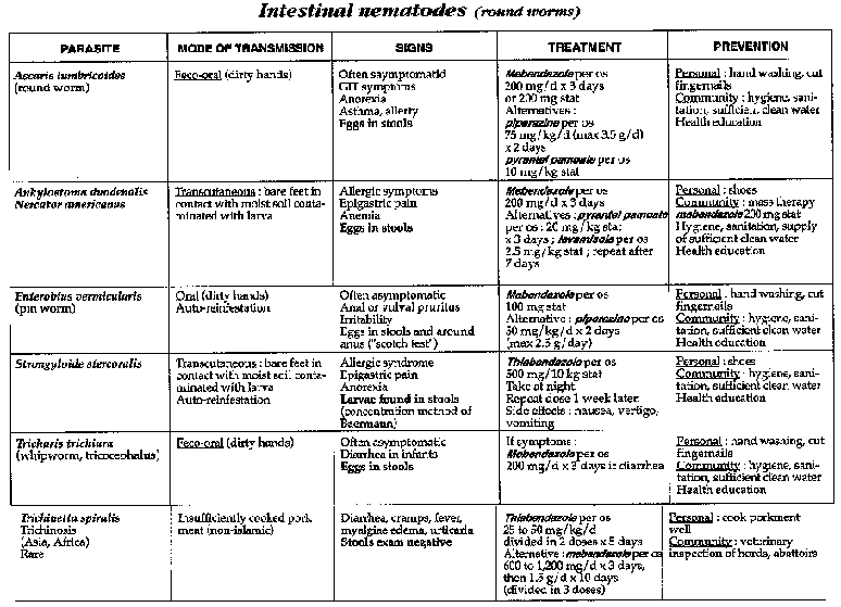
Table
Liver flukes
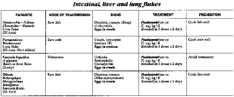
Table
Cestodes
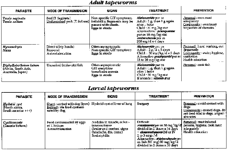
Table
Filariasis
Group of conditions caused by infection with various nematodes, the most common being Wuchereria bancrofti, Brugia malayi, Onchocerca volvulus, Loa loa and Dracunculus medinensis. Adult forms of both sexes live and reproduce in human lymphatics, in the skin or in deep tissues. Their larvae, microfilariae, reach the blood or skin and are thus the infective form for biting vectors as well as being the form upon which diagnosis is based.
Transmission is by vector: mosquitoes for lymphatic filariasis (Bancroftian and Malayan), blackflies for onchocerciasis, Chrysops flies for loiasis and tiny crustaceans (Cyclops) for dracunculiasis (Guinea worm).
Clinical features and diagnosis
See table.
Symptomatic treatment
- Inflammatory symptoms: acetylsalicylic acid (PO): 3 g/d divided in 3 doses or indomethacin (PO): 75 mg/d divided in 3 doses
- If allergic symptoms develop (e.g. urticaria, pruritis) promethazine (PO): 75-100 mg/d divided in 3-4 doses; child: 1 mg/kg/d divided in 3 doses or chlorpheniramine(PO):12 mg/d divided in 3 doses
Antiparasitic treatment
LOIASIS AND LYMPHATIC FILARIASIS
The main drug used is diethylcarbamazine, often abbreviated to DEC. It is essentially a microfilariacide and may not kill all adult worms. Therapy with DEC should always be supervised as the drug is often poorly tolerated (allergic reactions). Dosages should start low and be increased progressively. DEC is contraindicated during pregnancy. Usual presentation is in 50 mg tablets.
LYMPHATIC FILARIASIS
Adult: commence with 25 mg/d divided in 2 doses (= 1/8 tab x 2/d). Increase progressively by doubling the dose each day until the 5th day, dose is 200 mg x 2/d=2 tab x 2/d) x 10 days.Child: 3 mg/kg x 2/d x 10 days, to be reached progressively over 5 days.
A second therapeutical course can be repeated after 10 days.
LOIASIS
In this infection diethylcarbamazine can cause a fatal
encephalitis or allergic shock. Much care is needed. Reinfection after treatment
is very common, so if symptoms are mild, it may be better to withold therapy.
Dosage can be adjusted to extent of infestation
(beyond 50,000
microfilaria/ml blood: +++ caution).
Where treatment considered essential because of severity of infection: diethylcarbamazine
Adult: 3 mg x 2/d (= 1/32 tab x 2/d) the 1st day increasing progressively till in seven days 200 mg x 2/day (= 2 tab x 2/day) x 21 days.Child: begin progressively to reach in seven days 3 mg/kg x 2/d x 21 days.
Always give antihistamines in association.
If promethazine does not control reactions to treatment, treat with prednisone (or prednisolone): 15-30 mg/d in a single dose x 3-5 days, then decrease progressively. If necessary, dexamethasone IV or IM: 4-20 mg / kg.
Note: where Loiasis is endemic (West Africa), all treatment with diethylcarbamazine, should commence with 3 mg/kg x 2 days (protocole for Loiasis) whatever form of filaria is being treated. This is to avoid the sometimes fatal complications of inopportune treatment where there is also unrecognised associated loiasis.
ONCHOCERCOSIS
The treatment of choice is ivermectin (Mectizar), microfilaricide, 6 mg tablets.
|
Dose: |
150-200 |
micrograms/kg stat: |
| |
15-25 kg: |
1/2 tab |
| |
26-44 kg: |
1 tab |
| |
45-64 kg: |
1.5 tab |
| |
65-84 kg: |
2 tab |
The recommended long term management of communities is one dose every 6 months the first year, then a single dose annually.
Contraindications: child < 5 years, pregnant women, women in their first week of breast feeding.
Side efects are due to lysis of microfilaria (allergic manifestations, pain, fever) and respond well to antihistamines and acetyl salicylic acid. Rarely orthostatic hypotension occurs but responds to injected cofficosteroids (single dose or 1-2 days).
There is no problem with associated filarias even Loiasis.
If ivermectin not available: diethylcarbamazine in dosage for lymphatic filariosis.
TREATMENT OF MACROFILARICIDES
This should be abandonned as too dangerous.
Prevention Prophylaxis
- See table.
- Individual chemoprophylaxis for Loiasis is possible, 100 mg diethylcarbamazine PO/week in a single dose (or 2 doses of 50 mg/week). It is indicated for non residents going to an endemic zone provided, they are not already infected with Loiasis (risk of serious reactions).

Table
Malaria
Parasitic infection due to protozoa of the genus Plasmodium transmitted by the female anophylus mosquito. There are four plasmodial species: P. Falciparum, P. Vivax, P. Malariae, P. Ovale.
Clinical features
INCUBATION PERIOD
- 9 to 13 days for Falciparum.
- More than 15 days for the other three forms.
PRIMARY ATTACK
Continuous fever + malaise + headache +/- gastro-intestinal problems. Consider it in endemic zones.
SIMPLE MALARIAL ATTACK
Shivering, fever ("heat"), sweating, headache, bodyaches. Theoretically, every two days for Falciparum, Vivax, Ovale and every 3 days for Malariae
SERIOUS MALARIAL ATTACK
- Uniquely due to Falciparum
Occurs especially in the non-immune: new subjects, non-residents, children < 5 years, pregnant women, debilitated patients; or in subjects living in a zone of seasonal transmission.
- Associated, in varying degrees, with the following clinical signs
· Cerebral signs: mental clouding, coma (lasting more than 1/2 hour in children following a convulsion), convulsions (more than 2 times/24 heures in children in the age range for febrile convulsions), delirium, localising signs.
· Haemolysis (jaundice is rare in children), haemorrhagic syndrome (sometimes C.I.V.D.).
· Renal signs (rare in children) have bad prognosis: oliguria, anuria.
· Hypoglycaemia: especially in children and pregnant women.
· Pulmonary edema: especially in adults; almost always fatal.
· Hyperpyrexia: T > 40.5°C.
· Macroscopic haemoglobinuria.
CHRONIC MALARIA ("MALARIA CACHEXIA")
Due particularly to Falciparum, sometimes Vivax. Usually in children.
Associated with febrile episodes, severe anaemia with pancytopenia, wasting, constant splenomegaly.
Necessary to make repeated staned slides as the protozoa are less numerous.
Diagnosis
Diagnosis is made by presence of protozoa in the blood: thick and thin slides should be made in endemic zones for every fever > 38.5°C.
Note that blood films may be negative, even in a severe
attack
(pernicious) because of sequestration or parasites in the deep
capillaries.

Map
Principal antimalarials
- Quinine
· Tablets 100 to 500 mg
Indications: following IV
treatment or a final trial (except in pregnant women inzones of
multiresistance).
Side effects: tinnitus, rarely giddiness, nausea,
vomiting.
Not recommended for prophylaxis.
· Ampoules: available from 60 mg to 300 mg/ml. Never by IV,
either infusion or IM (although IM injection can be given in cases absolute
necessity, numerous complications can occur: sciatic nerve paralysis, muscle
necrosis, infection).
Indications: try to reserve quinine by injection for
serious cases of malaria.
No contraindications.
- Chloroquine(Nivaquine)
· Tablets 100 and 150 mg base
Indications: Vivax,
Malariae, Ovale and uncomplicated attacks of sensitive
Falciparum.
Side
effects: pruritis is common in black skinned patients
(non-allergic and
unresponsive to antihistamines), rarely gastro-intestinal disorders.
Used for
prophylaxis.
· Ampoules 40 and 50 mg base/ml. Never by IV, either IM or
SC or infusion.
Note: the doses for injection are weaker than oral
doses.
Indications: severe vomiting or serious chloroquine-sensitive
malarial. No contraindications.
- Amodiaquine(Flavoquine)
· Tablet 200 mg
Active against some
chloroquine-resistant varieties. Abandoned because of its toxicity
(agranulocytosis, hepatitis).
Must not be used for prophylaxis.
- Pyrimethamine-sulphadoxine(Fansidar)
· Tablet with 500 mg of sulphadoxine + 25 mg of pyrimethamine, orally
· Ampoule with 200 mg of sulphadoxine + 10 mg of
pyrimethamine/ml
(= 400 + 20/amp of 2 ml); IM (never IV)
Indications: treatment of simple attacks of Falciparum (in zones of intermediate resistance as 1st or 2nd choice.
Side effects: rare but serious: Lyell syndrome, Stevens-Johnson syndrome, agranulocytosis, especially when used for prophylaxis.
Contraindications: pregnancy or breast feeding, children < 2 years (avoid before 5 years).
Should be abandoned as prophylaxis. Not to be given in association with chloroquine (antagonism) or with mefloquine.
- Mefloquine (Lariam) (veryexpensive)
· Tablets 50 and 250 mg
Indications: simple attacks of multiresistant Falciparum (as 1st choice in zone III as 2nd or 3rd choice elsewhere).
Side effects: giddiness and digestive disturbances are common; rarely, acute psychosis, encephalopathy with convulsions, transitory but serious.
Contraindications: epilepsy, history of psychiatric disturbances, avoid in pregnant women.
Its use for prophylaxis is limited by its side effects.
- Halofantrine (Halfan) (very expensive)
· Tablet 250 mg
Tablets should be taken with a fatty
accompaniment
Indications: simple attacks of multiresistant Falciparum (as
1st choice in zone III, as 2nd or 3rd choice elsewhere)
Side effects: unobstrusive (pruritis, gastro-intestinal disturbances).
Contraindications: pregnancy or breast feeding.
Unusable for prophylaxis.
- Tetracyclin
· Tablet 250 mg
Indications: associated with quinine in areas where the Plasmodium
Falciparum is becoming less sensible to quinine (zone III), use only in severe malaria when the patient is able to swalow.
Side effects: nausea, vomiting,
photosensitization.
Contraindications: pregnancy and breast feeding, children
< 8 years old.
Cannot be used as prophylaxis.
- Doxycyclin
· Tablet 100 mg Indications, side effects,
contraindications: idem tetracyclin.
Usable for prophylaxis.
- Proguanil(Paludrine)
· Tablet 100 mg
Only usable for prophylaxis in
association with chloroquine including pregnant women and children.
Very few
side effects. No contraindications.
- Primaquine: gametocytocide
· Tablet 15 mg
Indications: avoidance of relapses of
Vivax and Ovale. Toxic: methaemoglobinaemia, haemolysis where G6PD deficiency
exists
(common in Africans,Asiatics and those of Mediterranean
origin).
Contraindications: pregnancy and in G6PD deficiency.
In practice,
few indications (expatriates leaving an endemic zone or where demanded by
national rules, especially as resistance to it has been described).
Drug resistance of P. Falciparum
RESISTANCE TO CHLOROQUINE
- Before speaking of resistance, verify:
· that treatment has in fact been taken,
· that the correct dose for weight has been prescribed,
· the absence of important diarrhoea and whether there has been vomiting within one hour of taking medication,
· the expiry date of the medication,
· that there has not been under-dosage due to confusion between the expression of the dosage as a chloroquine base and as a chloroquine salt(1).
- There must evidently be a Falciparum positive blood slide on
the first and third days of treatment, slides which have been theoretically
quantified. There is no chloroquine resistance with
P. Vivax, Malariae or
Ovale.
- If resistance is suspected, follow the recommendatios for the country concerned.
WHO has done in vivo testing which is rarely possible in routine practice.
- WHO classifies resistance to chloroquine into 3 types. Schematically:
· Early and late R 1: total disappearance followed by reappearance of the parasite
· R 2: noticeable fall without diseappearance of the parasite
· R 3: parasite level almost unchanged, indeed, increased
- Zones of resistance: see map. There is resistance to chloroquine almost everywhere where Falciparum rages and is more or less frequent depending on the country. Schematically, 3 zones can be distinguished:
· Zone I
Chloroquine sensitivity retained. Certain West
African countries, Central America except for Panama.
· Zone II
Spreading pockets of resistance, often of low
level (R1). East Africa, Northern India, part of West Africa.
· Zone III
Numerous chloroquine resistant areas of
raised level (R2-R3). Multiresistances. South East Asia, part of India and
Pakistan, Polynesia, South America.
RESISTANCE TO SULPHADOXINE-PYRIMETHAMINE
- Resistance to sulphadoxin-pyrimethamine (Fansidar), although less frequent, has for several years followed closely the distribution of chloroquine resistance. Very extensive in South East Asia and Brazil.
RESISTANCE TO QUININE AND MEFLOQUINE
- Resistance to quinine (type R1) and mefloquine, also exist principally in South East Asia.
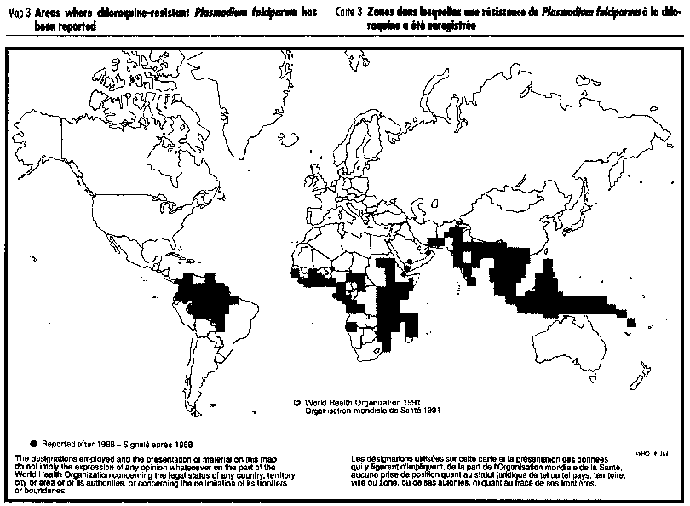
Map
Treatment of primary or uncomplicated attack
- Always use national guidelines. This is essential in countries where drug resistance occurs.
- For Vivax, Malariae, Ovale and for Falciparum in zone I
(chloroquine-sensitive):
chloroquine.
Adult: 600 mg base then 300 mg base at H6, D2 and D3
Child: 10 mg base/kg then 5 mg base/kg at H6, D2 and D3
Or a total of 1.5 g for an adult and 25 mg/kg for a child.
- For Falciparum in zone II (intermediate resistance)
· either chloroquine as first choice (eventually 10 mg/kg on D1, D2 then 5 mg/kg on D3, D4, D5) and in case of failure, sulphadoxine-pyrimethamine:
Adult: 3 tab in a single dose
Child: 1/2 tab/10 kg in a single dose
· or sulphadoxine-pyrimethamine as first choice. Theoretically, mefloquine or halofantrine are only used in cases of chloroquine and/or sulphadoxine-pyrimethamine failure, with quinine as the last recourse. In practice, account must be taken of national guidelines, availability and cost.
- For Falciparum in zone III
· either mefloquine
Adult and child: 15 mg/kg then
10mg/kg 8 hours later; repeat the dose if vomiting occurs less than one hour
after medicament taken.
In children, if the temperature is lowered before taking the medicine (antipyretics, cold bath), this will decrease the frequency of early vomiting.
· or (especially in pregnancy): quinine: 30 mg/kg/d divided
in 3 doses x 7 days
in association with tetracyclin:
Adult: 1.5-2g/d x 7 days
Child > 8 years: 50 mg/kg/d x 7 days
Tetracyclin is usually contraindicated in pregnancy or breast feeding and children < 8 years, but the vital risk serious malaria makes this a secondary consideration.
· or halofantrine (rarely available becaue of cost)
Adult: 3 doses of 500 mg at 6 hourly intervaals
Child: 3 doses of 8 mg/kg at 6 hourly intervals
It should be reserved for cases where resistance has been
proven.
The ingestion of halofantrine concurrently with fats doubles or
trebles its absorption.
Treatment of serious malaria
- Quinine as an infusion remains the treatment of choice in every zone. Commence with a loading dose of 20 mg/kg in an isotonic solution (if possible 5 % glucose) over 4 hours, then 10 mg/kg every 8 hours (or better, 4 hours with quinine, 4 hours through indwelling venous needle), until such time as the patient can swallow. Then change to oral medication for the remainder of the 7 days.
- SIf the patient has received quinine or mefloquine in the previous 24 hours without vomiting a loading dose will not be given (cardiac toxicity).
- If injection is not possible, use quinine IM in zones II or III in the same doses.
- In zone III, complete therapy with tetracycline.
- In zone I, chloroquine as IM (antero-lateral thigh) or SC can be used in place of quinine. Dosage: 3.5 mg base/kg every 6 hours or as an infusion: 10 mg/kg in an isotonic solution over 8 hours, then 5 mg/kg/8 hours. Transfer to oral therapy as soon as possible. The total dose should be 25 mg/kg.
COMPLEMENTARY TREATMENTS
- Convulsions: diazepam IV or IR (see "Convulsions"). For prevention, use with phenobarbital.
- Infusion: guard against excessive hydration if not sure of the integrity of renal function. Do not exceed 2,000-2,500 ml/days in adults and 40-50 ml/kg in children, except where there is presenting dehydration which needs to be corrected.
- Transfusion where there is profound or porrly tolerated anaemia (Hb < 5 mg/ml or haematocrit < 15 %). Check for HIV if possible.
- Safe position and nursing in case of coma, nasogastric feeding.
- Record urine output: if < 400 ml/day in adults or 12 ml/kg/day in children; regard patient as anuric. This normally requires fluid restriction. To avoid decreasing the perfusion rhythm and therefore the planned doses of quinine, attempt to initiate a diuresis with frusemide IV (ampoule 10 mg/ml, 2 ml) at the rate of 2 ampoules of 20 mg as required (do not exceed 250 mg in 100 ml of 5 % glucose administered over 20-30 minutes in adults; in children, the dose is 1 mg/kg/inj. repeated every 4-6 hours depending on the evolution).
- Hypoglycaemia: when an doubt, especially in children or pregnant women, give 20-50 ml of 30 % or 50 % glucose. Hypoglycaemia often relapses. In practice, in children, the perfusion of 80 ml/kg/day of 5 % glucose prevents the hypoglycaemia induced by quinine.
- OAP: frusemide.
- Antipyretics as necessary.
Specific case: treatment of a simple infection in a non-immune subject (expatriate) and in pregnant women
- Falciparum infection
· In sensitive zones: chloroquine.
either
Adult: 600 mg base then 300 mg base at H6, D2, D3, D4 and D5 (= 2.1 g)
Child:10 mg base/kg then 5 mg base/kg at H6, D2, D3, D4 and D5 (= 35 mg base/kg)
or
Adult: 500 mg base/ d x 5 days (= 2.5 g)
Child: 10 mg base/kg/d x 5 days (= 50 mg base/kg)
· In resistant zones
either mefloquine: the protocol is
the same as for protected subjects except for adults > 60 kg who must take
750 mg, 500 mg, 250 mg at 8 hourly intervals (= 3 tab, 2 tab, 1 tab).
or
halofantrine: the same protocol as for protected subjects.
· Pregnant women
- In sensitive zones: chloroquine
- In resistant zones:
quinine (follower by mefloquine if this fails)
- Vivax, Malariae or Ovale infections chloroquine
Prophylaxis
INDIVIDUAL
- Reserved for expatriates, and depending on the national protocol, for pregnant women.
- Depends on the region to which they are going: see the maps which follow.
- Depends on the length of stay, the season (transmission or not), the presence or absence of resistant Falciparum.
- Protection against anopheles:
These measures must assume
increasing importance:
· mosquito sprays impregnated with pyrethrinoid (permethrine or deltamethrine),
· long sleeves, long trousers and dark clothing at night,
· repellents,
· slow combustion anti mosquito coils.
- Information regarding prevention.
- Chemoprophylaxis: does not prevent infection with malaria but may avoid a serious attack. Commence the eve or day of departure and always continue for 6 weeks after return.
· In zone I
chloroquine: 100 mg base/day, 6 days each
week, or 300 mg in a single dose each week.
· In zone II
chloroquine: 100 mg base/day, 6 days each
week, in association with Proguanil 200 mg/day (in 1 or 2 doses).
· In zone III
Several possibilities depending on the
situation. There is no 100 % efficacious drug. nor any without side-effects.
either take mefloquine 250 mg (1 tab) 1 time/week only during the transmission season, or throughout the entire stay if this does not exceed 12 weeks; or where the stay does not exceed 1 month, take doxycyclin 100 mg (= 1 tab) each day, every day;
or take nothing, but carry at all times an effective treatment (mefloquine, halofantrine, quinine) to be taken if any symptoms suggestive of malaria appear.
Check with microscopic examination of the blood any time this is possible.
- Chemoprophylaxis incorrectly taken means lack of protections.
- Prophylaxis with sulphadoxine-pyrimethamine must be abandoned.
COLLECTIVE
It is necessary to carry further the battle against the anopheles mosquito, since mass chemoprophylaxis is no longer recommended (development of resistance, slowing or suppression of protection), except in pregnant women.
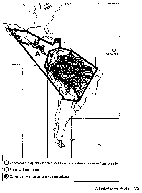
Map

Map
Trypanosomiasis
African trypanosomiasis = sleeping sickness
- African trypanosomiasis is caused by a flagellated protozoan, which is transmitted to humans by the tsetse fly (Glossina spp)
- There are two species of the parasite, each having a different geographical distribution:
· Trypanosoma brucei gambiense (West Africa)
· T.
b. rhodesiense (East Africa)
Clinical features
Clinical manifestations of infections with the two species are similar, except that T. b. rhodesiense infections tend to run a more rapid course.
- Primary stage: sometimes a painless chancre appears at site of bite. Incubation period very variable (days to years).
- Blood stage: fever, adenopathy, hepatosplenomegaly and facial edema. Presence of trypanosomes in blood and in lymph: gland puncture, blood film
- Cerebral stage: chronic meningoencephalomyelitis
· "Sleeping sickness": psychiatric, motor and sensory signs
· Disturbed sleep pattern: hepatosplenomegaly and adenopathy may resolve, blood film becomes negative for trypanosomes, specific serology positive, CSF (raised numbers of lymphocytes (> 5/mm3), raised protein, sometimes presence of trypanosomes, CATT test on serum, Elisa or CSF).
- Other manifestations: T. b. rhodesiense infections may be complicated by a fatal myocarditis.
Prevention
A trypanosomiasis control program must only be conducted in coordination with national health authorities. Consult specialized documents or monographs. Elements:
- Active case detection and treatment.
- Vector control.
- Notification of cases to health authorities (surveillance).
Treatment
The choice of regimen is based upon the results of CSF examination. If CSF is normal, the disease is considered to be in the blood stage. Abnormal CSF indicates cerebral involvement.
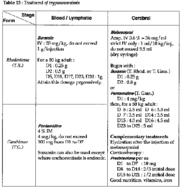
Table 13
Therapy should of course follow national guidelines. Refer also to the WHO monograph (Technical Report Series 739).
Where resistance to melarsoprol develops, use nifurtimox according to national guidelines or DFMO.
Ameriacan trypanosomiasis = Chagas' disease
Disease caused by Trypanosoma cruzi, transmitted to humans through the feces of infected reduviid bugs, which live in cracks in walls. T. cruzi infects humans via skin lesions (scratches, or bug bite) or mucus membranes, especially the conjunctivae.
Clinical features
- Incubation:10 to 20 days
- Acute phase:
· Chagoma: chancre, often on face
· Unilateral
edema of the eyelid and adenopathy
· Persistent fever, generalized
adenopathy
· Acute myocarditis: chest pain, CCF
·
Hepatosplenomegaly
· Meningoencephalitis: paralyses, convulsions
- Chronic phase (after a long latent period):
· Chronic cardiomyopathy: arrythmias, CCF, angina
·
Megacolon, megaesophagus
Diagnosis
- Acute phase:
· blood slide: often difficult to find the parasite.
· Xenodiagnosis: examination of the feces of reduvid bugs that have fed upon the patients blood.
- Chronic phase: serology.
Treatment (dispensary - hospital)
- In spite of progress, treatment for T. cruzi infections is not entirely satisfactory. The drug of choice is at present: nifurtimox(PO): 8 to 10 mg/kg/d divided in 3 doses x 3-4 months.
· No alcohol during therapy (Antabuse effect)
· Give prednisone ou prednisolone (PO): 1 to 2 mg/kg/day at the same time and taper off gradually.
NB: this use of corticosteroids is controversial: some sources
claim it can exacerbate the disease.
Side effects (may be severe): gastritis,
agitation, convulsions, tremor, paraesthesiae.
· Contraindications: pregnancy, history of convulsions.
- benzonidazole(PO): 5 to 8 mg/kg/day x 30 days.
· Side effects: rash, peripheral neuritis
Indications for therapy
- Both drugs are active during the acute phase.
Only
benzonidazole has an effect during the chronic phase.
- Give supportive treatment of convulsions, CCF and pain.
Prevention
- Mosquito nets.
- Vector control (insecticides): residual insecticides.
- Improved housing: plastered walls, corrugated iron rooves, cemented floors all reduce the vector habitat (thatch, small cracks in mud).
Leishmaniasis
Parasitic infection of humans and certain animal hosts caused by flagellate protozoans, Leishmania spp, transmitted by the bite of infected female Phlebotomus sandflies. Two major forms:
- Cutaneous and mucocutaneous leishmaniasis
· Old World, also known as oriental sore (also by many other local names). Occurs in the Middle East, Mediterranean, Ethiopia, India.
· New World, also known as espundia or mucocutaneous form, occurs in South America and Africa (Ethiopia...).
- Visceral leishmaniasis, or Kala-Azar
· Occurs in the Middle East, Mediterranean, India, East Africa, China, Latin America.
Clinical features
CUTANEOUS LEISHMANIASIS (ORIENTAL SORE)
AND MUCOCUTANEOUS
LEISHMANIASIS (ESPUNDIA)
- Incubation 2 to 4 months; single or multiple lesions appear on exposed areas of skin. Starts as a papule, which then extends in circumference and depth to form a crusty ulceration (dry form).
- Wet forms tend to evolve more quickly.
- Lesions tend to resolve spontaneously, leaving a scar.
- Lesion can extend to mucus membranes (mouth, nose, conjuntivae) and can be very mutilating (mucocutaneous form).
KALA-AZAR
- Persistent fever, pallor, anemia, weight loss, hepatomegaly, splenomegaly; sometimes adenopathy, diarrhea and hemorrhage.
- Raised ESR, raised gammaglobulins.
- If untreated, is invariably fatal.
- Serology: test for Kala-Azar (direct agglutination, clot Elisa. Always confirm by looking directly for parasites. Serology is of no value in cutaneous forms (false -, false +).
Diagnosis
- By indentification of Leishmania from skin lesions (cutaneous forms), or from blood, bone marrow, lymph nodes or spleen (kala-azar).
- May-Grunwald-Giemsa stain: parasites are intracellular and seen within histiocytes.
Treatment
- The main drugs are antimony compounds:
meglumine
antimonate(amp 5 ml = 1.5 g in IM): 50 mg/kg/d x 10 to 15 days
sodium
stibogluconate (amp. 1 ml = 100 mg in IM or IV):
|
Adult: |
6ml/day |
|
Child under 5 years: |
2 ml/day |
|
Child 5 to 14 years: |
4 ml/day |
Duration of 30 days, except with Indian kala-azar which is treated for 6 days only.
· Idiosyncratic reactions: fever, chills, cough, myalgia and rash. These reactions can be fatal so stop therapy if any of these symptoms appear.
· Therapy must be closely supervised as toxicity may appear late and is serious. Signs of toxicity are: fever, chills, cough, rash, polyneuritis, cardiac failure and renal failure.
- pentamidine (amp. 3 ml = 120 mg in IM): 2 to 4 mg/kg x 6 injections every 48 hours The patient should be supine during and after the injections as they can cause either hypoglycaemia or hyperglycaemia.
Indications (hospital)
- Cutaneous leishmaniasis
Both meglumine antimonate and
sodium stibogluconate promote healing but are not without danger.
· Systemic meglumine antimonate (course of IM injections) can be reserved for serious cases.
· For single lesions, or a small number of small lesions, local therapy can be tried instead. Give 1 to 3 ml of meglumine antimonate injected around and beneath the lesion, to be repeated if necessary.
- Visceral leishmaniasis
Either meglumine antimonate or sodium stibogluconate are given
systematically as described above. Strict supervision is necessary.
In case
of poor response or idiosyncratic drug reaction, use pentamidine.
Prevention
Vector control and, in some cases, control of animal reservoir.
|
| |||||||||||||||||||||||||||||||||||||||||||||||||||||||
Clinical Guidelines and Treatment Manual (MSF, 1993, 319 p.)
Chapter 7 - Bacterial infections
Meningitis
Acute inflammation of the meninges usually of bacterial origin with risk of progressing to encephalitis.
Clinical features
ADULT AND CHILD OLDER THAN 1 YEAR
- Classical meningeal syndrome with fever, meningism, neck stiffness, Budzinski and Kernig's signs positive: the patient lying extended, involuntarily flexes the knees when the neck is flexed or when the legs are raised vertically with the knees in extension.
- If severe: febrile coma, convulsions, localising signs, purpura fulminans.
CHILD UNDER 1 YEAR
Diagnosis much more difficult as classical meningeal signs are often missing. Always think of it in a sick child:
- refusal to eat, fever with diarrhoea, vomiting, drowsiness, plaintive crying, unusual behaviour;
- generalised or focal convulsions, coma;
- infant may be hypotonic, neck is often not stiff, fontanelle bulging even when not crying;
Localising signs:
- purpura may be minimal;
- slide tests negative;
- fever may be absent.
Differential diagnosis
Where malaria is endemic, it is vital to consider cerebral malaria (thick and thin slides).
Lumbar puncture
Do lumbar puncture whenever in doubt.
Cerebro spinal fluid (CSF) normal: clear, cells < 5/mm3, proteins < 0.40 g/1 (Pandy -).
In meningitis: polymorphs > 500/mm3, proteins = about 1 g/1 (Pandy +). CSF cloudy "rice water" = meningitis.
Whenever possible, ask for gram staining and direct microscopy for white blood cells.
Causative agents
MORE THAN 3 YEARS OLD
- Meningococcus (dry season)
- Pneumococcus (often linked with another focus: pneumonia, RTI)
- Rare other pathogens
2 MONTHS TO 3 YEARS OLD
- Haemophilus Influenzae
- Pneumococcus (often linked with another focus: pneumonia, RTI)
- Meningococcus (dry season)
- Rare other pathogens
LESS THAN 2 MONTHS OLD
E. Coli Listeria, Salmonella, Streptococcus B.
MENINGITIS OUTBREAK
Meningococcus A or C, mainly in Sahel areas, but sometimes elsewhere (Rwanda, Brazil). Outbreaks occur in dry season.
Antibiotic treatment in well equiped hospital
With the exception of oil based chloramphenicol IM, the antibiotic utilisable IV (chloramphenicol, ampicillin, penicillin) are short acting which necessitates IV injections every 6 hours. If this cannot be done then 1 injection every 8 hours. The important thing is that injections are given at regular intervals.
Choose the antibiotic effective against the invading pathogen.
MENINGOCOCCUS (GRAM-COCCUS)
- During an epidemic
The treatment of choice is oil based chloramphenicol IM, 1 single injection, to be repeated 24-48 hours later.
Dosage: 100 mg/kg/injection without exceeding 3 g/injection, giving half into each buttock according to the table.

Table
If clinical signs fail to resolve by 3rd day of oil based chloramphenicol, change to ampicillin IV.
If necessary, chloramphenicol per os can be used: 100 mg/kg/d divided in 3-4 doses x 7 days.
- When no epidemic
chloramphenicol IV
Adult: 5-6 g/day
Child: 100mg/kg/day
in 3-4 IV regularly spaced; change to oral treatment as soon as possible; total duration 7 days.
Other treatments are more expensive:
ampicillin IV
Adult: 10-12 g/day
Child: 200 mg/kg/day
in 3-4 IV regularly spaced; change to oral treatment as soon as
possible; total duration 7 days.
or
penicillin G IV
Adult: 20 MIU/day
Child: 200,000 IU/kg/day
in 3-4 IV regularly spaced; change to PPF IM at the same dose in 1 IM/d (it is not possible to change to oral penicillin V); total duration 7 days.
PNEUMOCOCCUS (GRAM + ENCAPSULATED DIPLOCOCCUS)
- ampicillin IVAdult: 10-12 g/day
Child: 200 mg/kg/day in 3-4 IV regularly spaced
or
- chloramphenicol IV
Adult: 5-6 g/day
Child: 100 mg/kg/day in 3-4 IV regularly spaced
In both cases, change to oral treatment as soon as possible; total duration 8-10 days.
HAEMOPHILUS INFLUENZAE (GRAM-BACILLI)
- chloramphenicol IV
Adult: 5-6 g/day
Child: 100 mg/kg/day in 3-4 IV regularly spaced
or
- ampicillin IV
Adult: 12-14 g/day
Child: 200-400 mg/kg/day in 3-4 IV regularly spaced
In both cases, change to oral treatment as soon as possible; total duration 8-10 days.
If lumbar puncture is not sterile on 3rd day, treatments can be combined. The chloramphenicol must be given 1 hour after the ampicillin, otherwise antagonism will occur.
Management in isolated conditions
- Lumbar puncture if in doubt. If CSF is cloudy, it is bacterial meningitis.
- Begin treatment without delay. Prognosis depends on the speed of initiating treatment.
· If available: oil based chloramphnicol IM (see doses above). Give 2 injections while preparing to transfer to a well equiped centre.
· If not, ampicillin IV or IM, ou chloramphenicol IV or IM, or penicillin IV or IM, or PPF IM. Give same dosage as for meningococcus.
Supportive therapy
-Ensure adequate nutrition and hydration (infusions, gastric tuve if necessary).
-Convulsions: diazepam IV or IR (see "convulsions").
-Coma: nursing +++ (care against bedsores, care of mouth and eyes).
-Purpura associated with shock: treat shock by restoring blood volume (see "Shock"), plus dexamethasone IV:
Adult: 16-20 mg
Child: 0,5 mg/kg
to be repeated as necessary.
Epidemic meningitis
-In risk zones (Sahel in dry season), check weekly incidence of meningitis.
-Decide the critical treshold at which point outbreak is considered an epidemic: either twice the usual weekly incidence (difficult to ascertain) or a level of 20 cases/100,000 inhabitants/weeks (20/100,000/week).
-Inform the local authorities in order to decide public health measures to be instituted.
-Identify the causative meningococcalagent (A or C) by a rapid agglutination test.
-Mass vaccination (vaccine anti A or anti A+C), with all its associated logistic problems, can be decided for the target population: 6 months to 15 years or 25 years. A single injection is sufficient to protect for 3 years. No contraindications. The vaccine is quite stable to heat.
-WHO advises against chemoprophylaxis (sulphonamides, rifampicine). For those who have been in contact with de disease: vaccination, information and supervision +++.
-Treatment: oil based chloramphenicol IM (see above).
Pertussis
Whooping cough is a childhood disease characterized by paroxysmal cough and tenacious sputum and caused by Bordetella pertussis. In developing countries it contributes to malnutrition.
Clinical features

Figure 3
- The cough can recur up to one year after the initial infection.
- Infants less than 3 months may develop apneic episodes or periods of hypoxia (cyanosis) without cough which may be fatal.
COMPLICATIONS
- Anorexia may precipitate protein-calorie malnutrition.
- Sub conjunctival hemorrhages, epistaxis, hemoptysis.
- Secondary infections of the upper and lower respiratory system.
- Encephalitis.
Treatment
(dispensary)
- Some authorities recommend antibiotic treatment during the prodromal (catarrhal) stage. This will not alter the course of the disease, but may reduce the period of infectivity and thus reduce transmission.
This is not practical except during epidemics, when all "colds" can be assumed to be prodromal pertussis.
erythromycin(PO): 50 mg/kg/d divided in 3 doses x 7
days
or
chloramphenicol(PO): 50 mg/kg/d divided in 3 doses x 7 days
- During the paroxysmal stage, antibiotics are of no use. Advise the mother to ensure adequate hydration, to humidify the air if possible, to remove the tenacious strands of sputum from the oropharynx, and, most important, to continue good nutrition, in spite of the child's anorexia and even if there is vomiting with each coughing spasm (feed the child again after the episode of vomiting).
(hospital)
- Secondary infections: antibiotics PO, IM or IV depending on severity:
ampicillin (PO): 100 mg/ kg/ d divided in 3 doses x 5-10
days
or
chloramphenicol(PO): 50 to 75 mg/kg/d divided in 3 doses x 5-10
days
or
cotrimoxazole (PO): 40 mg of SMX/kg/d divided in 2 doses x 5-10
days
- Infants less than 3 months of age should be admitted to hospital, if possible, and observed 24 hours a day.
Prevention
- Immunization (part of the routine program).
- During epidemics selective immunization of non-immune infants not manifesting clinical illness who have been in contact with pertussis cases.
Tetanus
Disease characterized by involuntary muscle spasms, usually fatal if untreated, caused by the tetanus bacillus. The portal of entry is either a wound, or in the case of neonates, the umbilical stump if this has been sectioned with a contaminated instrument. The tetanus victim is usually not immunized (importance of mass immunization programme).
Clinical picture
- Incubation period from 2 to 60 days following wound contamination.
- Trismus, muscle spasms, dysphagia.
- The least stimulus can incite paroxysmal muscle spasms.
Portals of entry
- Dirty wounds.
- Traditional practices during childbirth (e.g. circumcision, ear piercing).
- Surgery.
- Obstetric interventions; neonatal (2-14 days after delivery) and obstetric forms.
- Unsterile intramuscular injections.
Prevention (dispensary)
- Neonatal tetanus
· Sterile instruments for delivery and cutting of the cord.
· Training of and provision of equipment for traditional birth attendants.
· Immunization: 2 injections during pregnancy, the first as early as possible (during the first antenatal visit) and the second at least 1 month after the first and no later than 1 month before delivery.
- Routine immunization of all children (EPI).
- Correct wound toilet.
- Prophylaxis: often tetanus antiserum or booster doses of tetanus toxoid are not readily available. When they are, however, apply the following protocol:
· Patient fully immunized, having had a booster within the 10 previous years: no further treatment necessary.
· Patient fully immunized but last booster was more than 10 years ago: give single booster dose of tetanus toxoid (0.5 ml).
· Patient never fully immunized:
equine antitetanus
immunoglobulin 250 to 1,500 IU given SC. (Different sources give very different
doses.) At the same time start a full course (2 to 3 injections) of tetanus
toxoid.
-If serotherapy is not available, when there is a dirty wound in an non-immunized or inadequately immunised subject, clean and protect the wound, plus peni V or procaine penicillin in the usual doses for 5 days.
Treatement (hospital)
-Nurse the patient in a place with minimal sensory stimuli.
-IV fluids: maintain proper hydration.
-Nasogastric tube feeding.
-Equine tetanus immunoglobulin
Adult: 10,000 IU SC or IM. Start with a small challenge dose in case of allergic reactions.
Child: half the adult dose
Neonate: 1,500 IU SC or IM
Equine tetanus immunoglobulin is sometimes given intrathecally: e.g. for neonates, 1,500 IU via either lumbar or suboccipital puncture.
Reactions to equine tetanus immunoglobulin are treated with: dexamethasone (IV): 4 mg as needed.
-Penicillin G is given in order to eliminate any tetanus bacilli still releasing toxin in the wound:
penicillin G (IV): 100,000 IU/kg/d divided in 4 injections
-Control of muscle spasms:
diazepam (IV): 1 to 5 mg/kg/d by infusion
Further sedation if needed:
phenobarbitone (PO): 3 mg/kg/d divided in 2 doses by nasogastric tube.
Plague
- Zoonosis infecting many rodents and transmitted to man by fleas.
Plague was formally responsible for pandemics in Europe which caused high mortality.
- Large animal reservoirs persist: South-East Asia, Central Asia, East Africa, Madagascar, South America, USA. Human infection is becoming less common.
- Transmission to humans can be:
· direct, by the bite of an infected rodent,
· vector-bome (flea) from a rodent host. Both these modes of transmission give rise to sporadic cases only.
· Epidemics, however, arise when interhuman transmission occurs via flea vectors or, more important, by direct air-borne spread (pneumonic plague).
Clinical features
-Incubation
· 1 to 6 days for bubonic plague
· several hours to
2 days for pneumonic plague
-Bubonic form
High fever; painful buboes (adenitis) which are often inguinal (as fleas tend to bite the lower limbs) and produce a sero-sanguineous discharge. Without treatment the case fatality rate is high.
-Septicemic form
A rapidly fatal complication of the bubonic form.
-Pneumonic form
Severe pneumonia with hemoptysis, rapidly fatal. Highly contagious. Occurs either as a complication of the bubonic form or subsequent to primary air-borne pulmonary infection.
Diagnosis
-Identification of Yersinia pestis (staff must take great care not to innoculate themselves accidentally) by:
· aspiration of a bubo,
· sputum
examination,
· blood culture.
-Serology becomes positive early.
Treatment (hospital)
- Sulphonamides, streptomycin, tetracyclines and chloramphenicol but not the penicillins.
- Usual choice:
streptomycin (IM)
Adult: 500 mg every 4 hours for the first 2 days, then every 6 hours x 5-7 days
Child: 10 mg/kg every 4 hours for 2 days, then every 6 hours for 5 days
- If therapy is begun early the prognosis is good.
- chloramphenicol IV can also be used
60 mg/kg/d divided in
3-4 injections x 10 days
Prevention
- If cases are suspected, it is vital to:
· confirm the diagnosis bacteriologically,
· advise
local health authorities.
- Where possible plague patients should be isolated.
- Conduct concurrent and terminal disinfection of bedding, clothes...
- Take extreme care when handling exudates and cadavres.
- Give chemoprophylaxis for household contacts and health personnel during the entire period of exposure:
sulphonamides (PO): 40 mg/kg/d during period of
contact
or
tetracycline (PO): 20 mg/kg/d during period of contact
- Use appropriate insectides to control fleas.
- There is a plague vaccine which is effective for 6 months. Protection begins 7 days after vaccination.
- Long term prevention requires rat control, sanitation and good public hygiene.
Leptospirosis
- A zoonosis due to certain spirochetes also known as Weil's disease. In humans it may cause a febrile illness and acute hepatorenal failure.
- The main reservoir is animal usually rodents (especially the sewer rat), cattle, pigs, dogs, horses, wild animals.
- Infected rats, whether diseased or healthy carriers, excrete leptospirae in their urine and thus contaminate water and soil (bathing, poor hygiene and sewers...). The portal of entry being either mucous membranes, cuts or scratches on the skin.
Clinical features
-Incubation period of 7 to 10 days. Illness often biphasic. Fever, jaundice, meningism, proteinuria, hematuria and oliguria, hepatosplenomegaly, polyadenopathies.
-Can be associated with:
· Pulmonary symptoms: cough, pneumonia,
hemoptysis;
· diffuse haemorrhagic disorder: purpura, ecchymosis,
epistaxis...;
· severe renal insuficciency: oligo-anuria;
·
cardiac insufficiency (cardiac collapse).
-The meningeal signs may predominate (the CSF is macroscopically clear with raised lymphocytes and raised protein of 1 g/l). May progress to encephalitis.

Figure 4
Diagnosis aids
- WBC: frank leucocytosis excludes viral hepatitis.
- Urine: proteinuria, abundant pus cells, hematuria, and casts.
- Diagnosis is confirmed if spirochaetes are found in blood, urin or CSF; direct examination difficult; possible with fresh specimen using dark ground microscopy or very low light levels; otherwise Giemsa stain.
- Serodiagnosis: immunofluorescence or Elisa.
Treatment (hospital)
- Rest.
- Treat fever with paracetamol (not acetysalicylic acid, owing to risk of hemorrhagic disease).
- Antibiotics:
Must be commenced early in the illness if they
are to be effective:
Oral penicillin (or IV when serious neurological symptoms). Not IM because of risk of haematoma
Adult: 5-6 MIU / d divided in 3 doses x 7 days
Child: 100,000 IU/kg/d divided in 3 doses x 7 days
If allergic:
tetracycline (PO)
Adult: 1.5-2 g/d divided in 3 doses
Child > 8 years: 50 mg/kg/d divided in 3 doses
or
erythromycin: same doses as above x 7 jours
Prevention
- Rat control, sanitation and hygiene.
- Avoid swimming in endemic regions
- A vaccine exists for highly exposed individuals (farm workers etc).
Relapsing fever
Relapsing fever or borreliosis is a febrile illness caused by spirochaetes and transmitted to humans by lice or ticks. Immunity does not exceed 1 year.
There are two forms of the disease:
- Louse-borne caused by Borrelia recurrentis, worldwide distribution and transmitted by body lice. The reservoir is infected humans. The diseases tends to spread in epidemic fashion under conditions of crowding and hardship (e.g. war, refugees, cold and poor hygiene). Pockets notably in Ethiopia, Rwanda and Burundi.
Pockets notably in Ethiopia, Rwanda and Burundi.
- Tick-borne caused by many different strains of borrelia which are specific for each geographical region. Reservoir are humans but more importantly infected rodents. Spread by the bites of various ticks and therefore tends to present sporadically rather than epidemically.
Louse-borne fever is often associated with typhus (see Rickettsioses).
Clinical features
LOUSE-BORNE FEVER
Relapsing syndrome of fever, malaise, gastro-intestinal disturbances, jaundice, petechiae, meningism and hepatosplenomegaly.

Figure 5
Complications: hepatorenal syndrome, encephalitis, myocarditis, hemorrhage and miscarriage.
TICK-BORNE FEVER
Similar clinical picture.
Diagnosis
Thick and thin blood films during the fever peaks (Giemsa stain).
Treatment (dispensary - hospital)
- Same therapy for both forms: either tetracycline, chloramphenicol or erythromycin.
tetracycline (PO)
Adult: 1.5 g/d divided in 3 doses x 7 days
Child: 50 mg/ kg / d divided in 3 doses x 7 days
In severe disease, or < 8 years:
chloramphenicol
(PO)
75 ma/ kg/ d divided in 3 doses x 7 days
Single dose of doxycycline (PO) is also effective:
Adult: 200 mg
Child: 50 mg
For pregnant women:
penicillin V: 1.2 MIU/d divided in 3
doses x 7-10 days (beware of procaine penicillin: risk of haematoma).
- Therapy sometimes induces a Herxheimer reaction after the
first dose of antibiotics with fever, hypotension and neurological disturbances.
Therapy should therefore be supervised. Reactions are treated
with:
dexamethasone: 20 mg IM or IV
and/or
digoxine (IV) (see "Cardiac
failure")
The following regimen may reduce the chance of reaction:
Day
1: PPF(ou procain penicillin): 400,000 IU (IM)
Days 2 to 7: tetracycline (PO)
as above.
Prevention
LOUSE-BORNE FEVER
- Control of body lice: powder body and clothes with an
effective insecticide, usually:
1% lindane
If resistance, use:
malathion (powder) 1 %
or
propaxur
(Baygon)
or
deltamethine (K-othrine)
Use extreme care with these substances (toxic); handlers need instructions and supervision. Ask the MOH for guidance.
- In order to be effective the above measure should be applied to the entire population and repeated once after two weeks. This obviously requires good organization.
- Chemoprophylaxis:
doxycycline (PO): 200 mg/week in single
dose during epidemic.
Note that healthy carriers of relapsing fever are at risk of developing a Herxeimer reaction under chemoprophylaxis.
TICK-BORNE FEVER
Control of ticks: insecticides and personal protection.
Rickettsioses
- Group of diseases caused by Rickettsia spp, transmitted to humans by an arthropod vector.
- Transmission depends on the presence of:
· Reservoir of infection: human or animal.
· Vector: e.g. body lice, often associated with conditions of poor hygiene and sanitation.
· Crowding: such as in refugee camps.

Table 14
There are numerous other rickettsioses:
- scrub typhus
- Rocky Mountain spotted fever
- boutounneuse fever
- Q fever...
Their occurrence may be sporadic or epidemic.
Clinical features
-The different forms have a certain common core of clinical features
· high fever of sudden onset,
· severe headache,
chills, body pains,
· macular rash,
· prostration and coma.
-Evolution of the illness can be cyclical. After 2 weeks, there is a terminal crisis when the signs become more severe then resolve.
-Without therapy, grave and sometimes fatal complications may ensue: encephalitis, myocarditis and hemorrhagic disease.
Diagnosis
Confirmation can only be made by serology.
Treatment (hospital)
- Symptomatic for fever, dehydration... Not aspirin.
- Antibiotics:
tetracycline (PO)
Adult: 1-1.5 g/d divided in 3 doses x 7 days
Child: 50 mg/kg/d divided in 3 doses x 7 days
or
cloramphenicol (P O)
Adult: 2 g/d divided in 3 doses x 7 days
Child: 50 mg/kg/ d divided in 3 doses x 7 days
- Epidemic louse-borne typhus can be managed by giving a single oral dose of doxycycline: 200 mg (but risk of relapse).
- Therapy of typhus should not normally provoke a Herxheimer reaction (see louseborne relapsing fever) however, in some regions such as Ethiopia, the two diseases may sometimes co-exist in the same patient and a reaction is thus possible.
Prophylaxis
LOUSE-BORNE TYPHUS
- Control of body lice
· Powder body and clothes with an effective insecticide, usually 1% lindane.
· If resistance, use 1% malathion.
· Use extreme care with these substances: handlers need instructions and supervision. Ask the MOH for guidance.
· In order to be effective, the above measure should be applied to the entire population and repeated after two weeks. This obviously requires good organization.
- Chemoprophylaxis
doxycycline (PO): 200 mg/week in a single
dose during epidemic.
Risk of Herxheimer reaction in asymptomatic carriers of
relapsing fever.
FLEA-BORNE TYPHUS
Control of rats and fleas.
Brucellosis
- A systemic illness due to the gram negative Brucella. Transmitted to humans from infected cattle, sheep, goats or pigs, either by direct contact with infected tissues or by ingestion of milk.
- Often underdiagnosed, it is probably frequent among animal herders. It tends to occur predominantly among young males, who often have most contact with animals.
Clinical features
- Incubation period from 5 to 30 days.
- Acute brucellosis with septicemia
Oscillating fever,
sweats, flitting pains in the bones, joints and muscles.
Fever then plateaus
at 39 to 40°C, with tachycardia.
Defervescence after 10 to 14
days.
Hepatosplenomegaly and generalized adenopathy occur often with a group
of nodes gathered around a single larger one.
- Subacute brucellosis with focalization
Localized foci of
infection that persist and evolve autonomously. But mainly osteoarticular
(sternocostal, knee, tibia, spine, sacro-iliac).
Also meningeal and
encephalitic foci occur.
Note: brucellosis can minic Pott's disease, osteitis
or tuberculosis meningitis.
- Chronic brucellosis
Low grade fever, fatigue, vague pains
and sometimes infectious foci (such as arthritis).
Diagnosis
- Leucopenia with relative lymphocytosis
- Reaction to intradermal antigen
- Serology: rising titres in Wright's haemagglutination test or the Rose Bengal card test.
Treatment
ANTIBIOTICS
- Tetracycline (PO)
Adult: 2-3 g/ d divided in 3 doses
Child > 8 years: 50 mg/kg/d divided in 3 doses
- Cotrimoxazole (PO): tab 400 mg of SMX + 80 mg of TMP
Adult: 6 cp/ d divided in 2 doses
Child: 60-70 mg of SMX/kg/d (or 15 mg of TMP/kg/d) divided in 2 doses
- Streptomycin(IM)
Adult: 1 g/d in 1 injection
Child: 15 mg/kg/d in 1 injection
- Rifampicine (PO)
Adult: 900 mg/d in 1 dose
Child: 20 mg/kg/d in 1 dose
- Doxycycline and chloramphenicol also effective. Streptomycin can be replaced by gentamicin. Never use streptomycin or rifampicine alone.
RECOMMENDED MANAGEMENT
1. First treatment: tetracyclines x 6 weeks + streptomycin for
first 3 weeks.
2. Second treatment: cotrimoxazole for 2-3 months.
3. Third
treatment: tetracyclines+ rifampicine x 45 days.
INDICATIONS
- Acute brucellosis
Use first treatment.
- Brucellosis affecting bones
Use first treatment but
continue tetracyclines a further 45 days or better:
tetracyclines+
rifampicine for 3 months when possible.
- Neurologic attack
Add rifampicine to combined
tetracyclines-streptomycin.
- Pregnancy, breast feeding or children < 8 years
Use
cotrimaxazole, or rifampicine + streptomycin (if illness does not resolve).
- Relapse
Use first treatment if not already tried. If used
before, add rifampicine or change to cotrimoxazole (never use cotrimoxazole and
rifampicine together: antagonism).
- Chronic brucellosis
Only give antibiotic therapy if
persistent focus of infection, otherwise only analysis.
Prophylaxis
- Veterinary measures.
- Washing of hands and clothes after
contact with animals.
- Boil milk, avoid fresh cheeses and partially cooked
meat in endemic
zones.
Typhoid fever
Systemic illness caused by Salmonella typhi with foci of infection in the lymph and intestine. Transmitted either directly (unwashed hands) or indirectly (contaminated food or water).
Clinical features
- High fever, severe headache, insomnia, prostration, epistaxis.
- Either diarrhea or constipation, abdominal pain, bloated abdomen
- Splenomegaly, rose spots, pulse not in accord with fever.
- Complications (which may appear even during convalescence under therapy): GIT perforation or hemorrhage, peritonitis, septicemia, myocarditis, encephalitis.
- Leucopenia.
- Widal test (serology) becomes positive around the 8-lOth day (for the O antigen non-specific test).
- S. typhi can be isolated from blood or stool during the first two weeks.
Treatment (hospital)
- Close observation for complications.
- Treat fever and hydrate.
- Oral antibiotics are more effective than IV or IM (since the focus of infection is in the lymph nodes of the small intestine).
· First choice
chloramphenicol (PO)
Adult: 2 g/d divided in 3-4 doses
Child: 75 to 100 mg/kg/d divided in 3-4 doses
Start initially with half the dose the first day, and increase progressively.
· Alternatives (if resistance or contra-indication to
chloramphenicol):
ampicillin (PO) (progressive dose)
Adult: 4 to 6 g/ d divided in 3-4 doses
Child: 100 mg/kg/d divided in 3-4 doses
cotrimoxazole (PO) (1/2 dose and increase progressively over 3-4 doses)
Adult: 1,600 mg of SMX / d divided in 2 doses
Child: 40 mg of SMX/kg/d divided in 2 doses
- If patient cannot take antibiotics by mouth give IV initially but change to oral route as soon as possible.
- Continue treatment for 2 weeks after patient is apyrexial.
Prevention
- Isolation of cases
- Disinfection of excrete with chlorine
solution 2% or cresol 4%.
- Personal hygiene: hand washing and careful food
preparation.
- Community hygiene: water, sanitation and health
education.
|
| ||||||||||||||||||||||||||||||||||||||||
Clinical Guidelines and Treatment Manual (MSF, 1993, 319 p.)
Chapter 8 - Viral infections
Measles
- Also called rubeola and morbilli, it is one of the commonest childhood infectious exanthems. Among children in developing countries it is a serious illness with high mortality, especially when associated with malnutrition. Measles often precipitates acute malnutrition. Prevention by universal immunization of young children must always be a high priority.
- Measles is never subclinical, however recent studies have shown that the severity of the disease is related to the infective dose of virus. Crowding tends to increase mortality.
Clinical features

Figure 6
Complications
These must be looked for in all patients.
- Serious signs: persistent fever with darkening of the rash ("black measles") and subsequent desquamation.
- Stomatitis: compromises sucking and eating.
- Laryngitis: distinguish a benign prodromal laryngitis from that due to a secondary infection, which may be severe.
- Croup and otitis media.
- Bronchopneumonia: usually severe; gram negatives or staphylococcus.
- Diarrhea: either due to virus or from a secondary infection.
- Vitamin A deficiency: keratoconjunctivitis. Measles increases the con-sumption of vitamin A and often precipitates xerophthalmia.
- Encephalitis: caused by the measles virus itself; it occurs on about the 5th day of the rash.
- Malnutrition: precipitated by anorexia, stomatitis, fever, vomiting, diarrhea and other complications. Also important are frequent harmful cultural taboos that impose fasting upon a child with measles.
Treatment
(dispensary)
- Active case-finding during epidemic, if practical (home visits).
- Treat the fever.
- Keep well hydrated.
- Observe closely for complications.
- Give prophylaxis against conjunctivitis: drops or ointment.
- Give prophylaxis against xerophthalmia: vitamin A
|
Infants: |
100,000 IU in single dose on day 1, day 2 and day 8 |
|
After 1 year: |
200,000 IU in single dose on day 1, day 2 and day 8 |
- Encourage good oral hygiene.
- Maintain adequate protein-calorie intake: educate mothers (especially if cultural taboos against feeding exist), continue breast feeding, provide supplementary feeding if available (but do not admit to a feeding center until after infectious period).
- Antibiotics are often given prophylactically:
penicillin V
(PO): 100,000 IU/kg/d divided in 3 doses x 5 days
or
cotrimoxazole (PO);
60 mg of SMX/kg/d divided in 2 doses x 5 days
(dispensary - hospital)
- Treat secondary infections with antibiotics:
ampicillin: 100 mg/kg/d divided in 3 doses or (per os, IM or IV according chloramphenicol: 75 mg/kg/d divided in 3 doses to gravity x 7-10 days) or cotrimoxazole: 60 mg of SMX/kg/d divided in 2 doses
-Give supportive therapy for meningoencephalitis:
Adequate
hydration, good nursing, nasogastric feeding and control convulsions with
diazepam.
Prevention
- Education of mothers must be part of the MCH program.
- Immunization:
· A single injection gives good protection. Ideally should be given at the age of 9 months, but is often given later.
· Measles immunization is one of the highest priorities in refugee settings and other situations where crowding, poor hygiene and precarious nutritional status combine to encourage both transmission and the emergence of complications.
· There is an Oxfam/WHO measles immunization kit that is designed for emergency situations. Newly arrived refugee populations should be immunized during the first days of the emergency and all new arrivals should be immunized on entering. The target age-group is children from 9 months to 12 years (up to 5 years if resources are very scarce).
Poliomyelitis
- Acute infection due to the three strains of poliovirus affecting infants and young adults. It is endemic in developing countries but may occur in seasonal epidemics among non-immune persons. Transmission is feco-oral.
- Polio should disappear when immunization is universal. A very high proportion of cases are asymptomatic and these healthy carriers are the reservoir of infection: only 0.1% of infections give rise to paralysis. Polio occurs endemically in developing countries but seasonal epidemics can also occur.
Clinical features
- Febrile flu-like illness, often with diarrhea.
- Sometimes aseptic meningitis.
- Paralytic forms: paralysis is of sudden onset (often noted on waking in the morning), asymmetrical and hypotonic. It is associated with fever, hyporeflexia, risk of respiratory paralysis and eventual muscle wasting.
Treatment (hospital)
Is supportive only:
- Treatment of the fever and diarrhea.
- Rest, nursing care
for paralytic cases.
- Physiotherapy once signs have stabilized.
Prevention
- Immunization with live attenuated trivalent oral polio
vaccine
(OPV) with first dose at birth (new WHO recommendation) and then 3
more doses at the 6th, 10th and 14th weeks.
- Salk injectable killed vaccine at the 6th, 10th and 14th weeks.
- Theorically, boosters 1 year later and 5 years after that.
Public health measures
Community-level management:
- A paralytic case signifies that the virus is in circulation.
- Confirm the diagnosis serologically (rising titres).
- The immunization status of the community should be determined: date of the most recent program, coverage...
- If necessary, plan and implement a mass immunization program for children aged from 3 months to 5 years and carry out an "outbreak investigation".
Arbovirus diseases
Viral illnesses that usually have an animal reservoir and are trans-mitted to humans by mosquito vectors. They occur either sporadically or in epidemics.
Clinical features
Different viruses have different manifestations; however there are four main syndromes:
- Flu-like viral illness: e.g. dengue fever (SE Asia and the Pacific).
- Encephalitis: fever, neurological signs, convulsions, coma, sometimes meningism with a clear CSF. E.g. Japanese B encephalitis (SE Asia).
- Hepatorenal syndrome: fever, jaundice, oliguria, albuminuria, sometimes hemorrhages. E.g. yellow fever (West Africa, South America).
- Hemorrhagic fever: clinical picture of severe dengue fever plus shock and hemorrhagic manifestations in skin and mucosae. E.g. dengue hemorrhagic fever (usually only seen in children in SE Asia); also diseases such as Lassa fever, Ebola and Marburg virus, which have caused fulminant and deadly epidemics in Africa.
Treatment (hospital)
There is no causal therapy. Treatment is supportive.
Prevention
- Immunization: only a vaccine for yellow fever is available. A single injection protects for 10 years.
- Personal protection: mosquito nets, adequate clothing.
- Vector control: sanitation, destruction or management of vector breeding sites (e.g domestic refuse, water containers for urban AEdes aegypti, the vector of yellow fever).
- Epidemic management:
· Confirm the diagnosis: virological and serological studies in the nearest reference laboratory. One should collect detailed clinical and epidemiological information.
· Alert the local authorities.
· Plan control measures with the local authorities: vector control, public education and an immunization campaign.
Rabies
Viral zoonosis transmitted to humans in the saliva of an
infected animal.
Innoculation can be by:
- bite: dog, cat, wild animal, vampire bats (South
America),
- licking open skin lesions: cats, dogs, goats...
Principles of preventive tberapy
- The risk of exposure to rabies is higher in developing countries because of the high prevalence of the disease in stray animals.
- Clinical rabies in humans is invariably fatal but can be prevented with vaccine and antiserum after exposure.
- The incubation period in humans is from 2 weeks to several months depending on the severity and site of innoculation.
- An infected animal sheds rabies virus in its saliva before it develops signs of disease (14 days for dogs and cats).
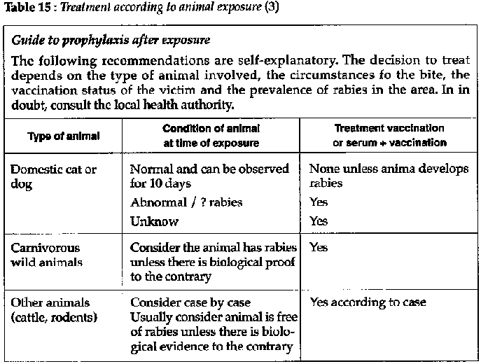
Table 15
- There are two types of exposure:
· Benign: contact of saliva with scratches on the skin; minor bites on the trunk or proximal limbs.
· Serious: contact of saliva with mucus membranes; bites on the face, head, neck, hands, feet, genitals; and bites from a wild animal.
Management of a person exposed to rabies (dispensary)
AFTER BITE
- Wash wound with soap, rinse, then dry thoroughly.
- Clean wound with chlorhexidine-cetrimide or other antiseptic and do not suture.
- Give tetanus prophylaxis.
- Capture and observe the animal for 15 days.
- Treatment:
· antirabies serum (Pasteur): prepared from horse
immunoglobin.
· rabies vaccine (Pasteur): human diploid cell vaccine
(HDCV).
- Note: the old vaccines (e.g. duck embryo) require several injections (7-14) and have allergic and neurological complications.
INDICATIONS FOR VACCINATION
- Depends on the condition of the animal at the time of the bite and after 15 days.
- There are two regimens:
· Benign exposure: simple rabies vaccination 1 dose (SC or IM) on day O (the start of the schedule), day 3, day 7, day 14, day 30 and day 90.
· Serious exposure: serum + vaccination Day O (as soon as possible after the bite): give serum 20-40 IU/kg IM Then Day 1, 8, 15, 30 and 90: give 1 dose of rabies vaccine.
Preventive measures for exposed personnel
Personnel who may be exposed to rabies (e.g. veterinarians, technicians) should be given 3 doses of rabies vaccine (HDCV) on days 1, 7, 21 or 28 and a booster dose after 6 months.

Table
16
Hepatitis
Several viral infections come under the heading viral hepatitis, each having its own epidemiology, clinical characteristics, immunology and prognosis. Hepatitis A, B, D and E occur in the tropics. The geographic distribution of hepatitis C is not yet known. All hepatitis, when they resolve, result in life long immunity, but not shared immunity.
The old terminology, non A-non B (A like-B like) has now been changed to hepatitis C and E. The "defective" virus D needs the presence of virus B to develop.
Principle characteristics are summarized in table 17.
Clinical features
ACUTE HEPATITIS
Nausea, fever, fatigue, abdominal discomfort, followed by the appearance of jaundice having an element of biliary obstruction, dark urine and stools more or less pale.
SUBCLINICAL INFECTION
Mild or anicteric infection is the most common but exposes the sufferer to the same risks.
FULMINANT HEPATITIS
Severe acute infection that leads to necrosis and liver failure. It is associated with high mortality.
CHRONIC ACTIVE HEPATITIS
May lead to cirrhosis and eventually hepatoma.
Treatment
- Symptomatic: rest, caution in prescribing analgesics (ea. acetyl salicylic acid, paracetamol), correct but not specific diet and hydration.
- Avoidance of corticosteroid therapy. Several medications are contraindicated.
Vaccination
Plan to include anti-B vaccine in the Expanded Program of Immunization (EPI).
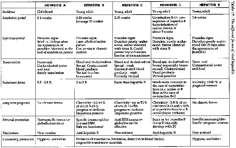
Table
17
A.I.D.S. and infection by VIH
AIDS, or Acquired Immune Deficiency Syndrome,is the most serious form, the end stage of infection by HIV (Human Immune-deficiency Virus). The virus attacks the immune system by infecting and then destroying the T4 lymphocytes.
Infection by HIV develops as a function of time,
schematically:
- Incubation period
From infection by the virus to the appearance of specific anti-HIV antibodies, lasts on average 6 weeks, sometimes marked by a non specific febrile syndrome (pseudo influenza syndrome).
- Asymptomatic period
This is the seropositive phase which can last years. The diagnosis depends on the detection of specific anti-HIV antibodies in the blood. On average, 50 % of seropositives progress to AIDS in 10 years (with actual decline).
- Symptomatic period
The immune deficiency syndrome is manifested clinically, it is the clinical AIDS phase. The pre-AIDS syndrome which was at a certain time defined by the term ARC (AIDS-related complex), is les and less recognised as a physiopathological entity. This is why there is no mention made of it here.
Epidemiology
HIV/AIDS infection is pandemic, occurring in epidemic form on 5 continents: however, not all populations are uniformly affected in one country or another or in the midst of the same country.
PREVALENCES
- Seropositives
In April 1991, WHO estimated that 8-10 million adults in the world had been infected by the virus since the beginning of the epidemic. Of these more 60 % were in Sub-Saharan Africa and around 1 million were children.
- Clinical AIDS
In April 1991, WHO estimated the number of clinical AIDS cases to be 1.5 million since the beginning of the epidemic (345,000 notified cases), 500,000 cases were children.
- Serotypes
Two serotypes have been identified: HIV 1 and HIV
2.
HIV 1 is the most widespread.
HIV 2 is particularly found in West
Africa and spreads less rapidly than HIV 1. However, the modes of transmission
and the clinica1 picture do not differ from one serotype to the other.
HIV TRANSMISSION
- Sexual transmission
During improtected homosexual and heterosexual relations. 70 % of the world's HIV infections result from heterosexual transmission.
- Blood transmission
By transfusions, contaminated surgical instruments, contaminated syringes and needles, by contact of even a minimal wound with contaminated blood (surgery or childbirth delivery without gloves, injury or prick with a needle or instruments contaminated with infected blood).
- Materno-faetal transmission
During pregnancy or delivery.
Note: transmission breast feeding is possible: nevertheless, the relationship between benefits and risks is such tnat WHO continues to recommend this form of feeding.
- HIV is not transmitted by saliva, mosquitoes, air, water, food, skin contacts, clothing, cooking utensils, the movement of general daily life.

Table 18
Projections
- Groups at risk: heterosexual transmission is the origin of 70 % of cases of infection in 1991, the groups at risk for the decade 1990-2000 will be heterosexual populations with multiple partners.
- Geography: 90 % of cases will survive in developing countries, Sub-Saharan Africa, Asia and Latin America.
Clinic factors
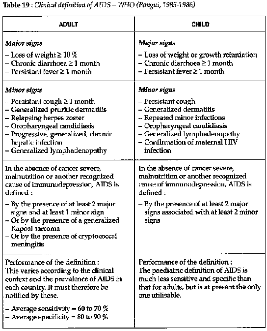
Table 19
This exclusively clinical definition is intended for developing countries lacking laboratory facilities (culture and/or histology). It is, above all, an indispensable tool in the surveillance of AIDS (notification of cases) and an elaborate clinical tool permitting clinicians to make a diagnosis with the maximum precision.
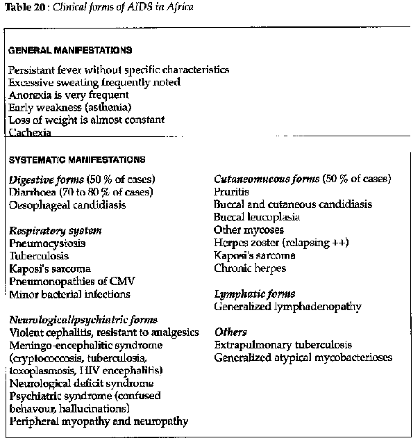
Table 20
Serological diagnosis of HlV infection/AIDS
In conditions in the field, the diagnosis of HIV infection in the asymptomatic adult can only the serological, i.e. the presence of specific anti-HIV antibodies in the blood is the sign of infection.
SEROLOGICAL TESTS

Table 21
In practice, the serological diagnosis of suspected AIDS is of no therapeutic interest. It can be justified for certain suggestive clinical tables of AIDS, but without satisfying the criteria of WHO clinical definition.
PRINCIPLES OF SEROLOGICAL DIAGNOSIS
To prescribe a serological test and announce the result negative or positive, for an HIV test, it must be combined with all the following conditions:
1. The patient must have received appropriate information about the consequences of a positive result and have given prior permission to be tested for anti-HIV antibodies.
2. Positive results from a blood sample must be confirmed by Western-Blott.
3. The seropositive individual must be able to benefit from further medical care and advice +++.
4. Confidentiality.
Treatment of HIV infection and AIDS
ANTIRETROVIRAL THERAPY
The only molecule in current commercial production is AZT (Azidothymidine). Its cost (2,000 - 3,000 US$/year) and the necessary techniques for therapy prevent, at present, its general use in developing countries.
TREATMENT OF OPPORTUNIST OR ASSOCIATED INFECTIONS (WHO GUIDELINES)
See tables 22a, 22b and 22c.

Table 22a

Table 22b
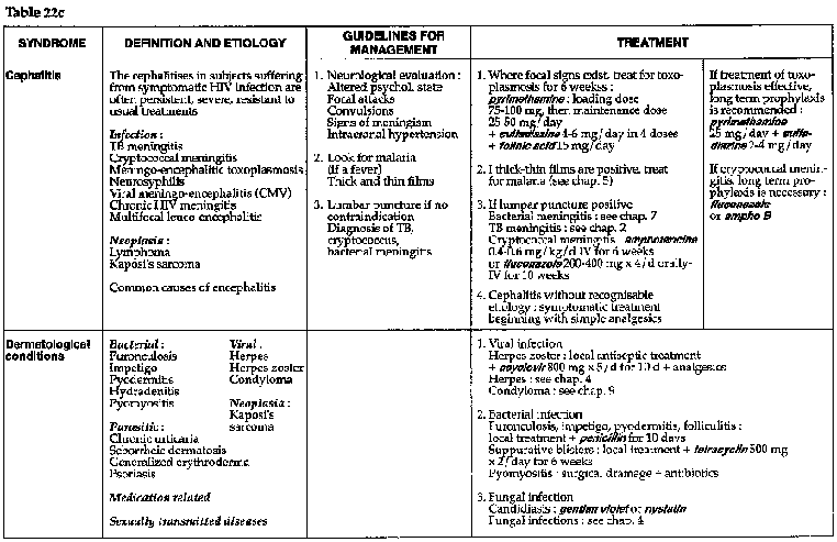
Table 22c
Prevention of transmission of HIV infection and AIDS
DURING SEXUAL INTERCOURSE
Systematic use of condoms is the only reliable prevention.
DURING TRANSFUSION
Strict respect for the indications for transfusion and systematic serological sampling of blood donors constitute the essential principals for the safety of transfusions.
See M.S.F. practical guide: "Practical transfusion in isolated surroundings - prevention of transmission of HIV"
IN MEDICAL FACILITIES
The prevention of the transmissin of HIV infection in the course of treatment takes place by reinforcement and strict respect for classical measures of hygiene:
- correct sterilization and disinfection of medical
material,
- avoidance of injections which are not strictly necessary,
-
precautions to avoid accidental contamination with soiled instruments,
-
precautions to avoid contact with potentially infected biological liquids.
See M.S.F. practical guide: "Recommendafions to prevent HIV transmission in health care facilities in developing countries "
|
| ||||||||||||||||||||||||||||||||||||||||||||||||||||||||||||||||||||||||||||||||
Clinical Guidelines and Treatment Manual (MSF, 1993, 319 p.)
Chapter 9 - Other conditions
Cardiac failure
Syndrome characterized by the failure of the myocardium to maintain an adequate cardiac output. Often called congestive cardiac failure, or CCF.
Clinical features
- Exertional and paroxysmal nocturnal dyspnea (pulmonary
edema).
- Hepatomegaly (tender liver on palpation).
- Ankle edema.
-
Tachycardia with gallop rhythm.
- Basal crepitations on auscultation of both
lung fields.
There are 3 forms of cardiac failure:
- Left ventricular failure
· Dyspnea: either exertional, recumbent (as in paroxysmal nocturnal dyspnea), or fulminant (acute pulmonary edema).
· Crepitations (rales) in the lung bases on auscultation (may be absent in infants); sometimes pleural effusion.
· Tachycardia, gallop rhythm.
- Right ventricular failure
· Edema: especially of the ankles and lower legs.
·
large, tender, sometimes pulsatile liver.
· Raised jugular venous
pressure.
- Biventricular failure: combination of right and left sided signs.
Symptomatic treatment (hospital)
- Half-sitting position, oxygen if available.
- Exclude salt
from diet.
- Drain any pleural effusion.
- Diuretics:
· Acute
pulmonary edema
furosemide (IV)
Adult: 20 to 40 mg/IV, repeated as needed
Child: 1 mg/kg/IV, repeated as needed
Compensated cardiac failure
furosemide (PO)
Adult: 20 mg/ d divided in 2 doses
Child: 1 to 2 mg/kg/d
Furosemide therapy depletes potassium and therefore the patient
should be supplemented:
potassium chloride: 1 g/day, 5 days out of 7
If furosemide is ineffective, use an aldosterone
antagonist:
spironolactone (PO)
Adult: 100-200 mg/day in single dose
Child: 3 mg/kg/day
· If furosemide or spironolactone are not immediately effective in acute pulmonary edema, two measures can reduce the load on the failing myocardium:
- Rotating tourniquets
- Venesection: bleed 200 to 400 ml; ensure first that significant anemia is not contributing to the cardiac failure.
- For left ventricular failure only
· In urgent situations (e.g. acute failure) digoxin (IV)
|
Adult: |
loading: 0.25 mg/injection, 3-4 injections in first 24 hours |
| |
maintenance: 0.25 mg/24 hours in 1 injection |
|
Child: |
loading: 0.010 mg/kg/injection, 3-4 injections in first 24 hours |
| |
maintenance: 0.010 mg/kg/24 hours in 1 injection |
In non urgent situations digoxin (PO)
|
Adult: |
loading: 0.5 -1 mg/d divided in 2-3 doses x 2-3 days |
| |
maintenance: 0.25 mg/ d, for 5 days out of 7 |
|
Child: |
loading: 0.015 mg/kg/dose: 3-4 doses/24 hours x 2-3 days |
| |
maintenance: 0.015 mg/kg/d in a single dose, for 5 days out of 7 |
- Treatment should be supervised: weight, dyspnea.
- Complications of treatment are bradycardia, arrhythmias and embolism.
- Pneumonia sometimes precipitates or complicates CCF. Treat with appropriate antibiotics.
Always seek a treatable cause.
The regimens below will usually be supplemented by symptomatic therapy of the CCF.
- Anemia: if severe enough to cause CCF may need transfusion. Great care is needed because of the danger of fluid overload: usually furosemide is given at the same time as any transfusion.
- Beri-beri: think of this, especially in SE Asia thiamine (vitamin B1)
Adult:200 mg IM or IV/day
Child:50 to 100 mg IM or IV
Continue with at least 200 mg / day PO for several weeks.
- Endocarditis
penicillin G (IV): 100,000 IU/kg
+
gentamicin (IM): 3 mg/kg/day
- Chagas'disease.
Acute rheumatic fever: myocarditis may lead to CCF in the acute
stage.
penicillin G or PPF (IM): 100,000 IU/kg/d x 10 days.
prednisolone
(PO): 2 mg/kg/d x 3-5 days, then decreasing dose regimen over 7 to 10 days.
Prophylaxis:
benzathine penicillin (IM): 1.2 - 2.4 MIU every
2 to 4 weeks.
< 15 years: 1.2 MIU every 2 weeks
> 15 years: 2.4 MIU
every 2 weeks
for several months if
possible.
Hypertension
- Before diagnosing hypertension, the BP must be checked several times with the subject resting.
- Drug therapy should only be instuted for BP consistently above 160/90 mm Hg (or 140/9Ofor pregnant women).
- Therapy must be closely supervised, otherwise side effects can be serious.
Treatment
ESSENTIAL HYPERTENSION (dispensary)
No evident cause, in a non-pregnant subject.
- Low-salt diet: follow-up one week later.
- If BP still > 160/90: drug therapy.
hydrochlorthiazide
(PO): 50 mg/ d, best taken in the morning.
Give potassium supplement (e.g.
advise bananas in diet).
- If no improvement after one week give in
addition:
methyldopa (PO): commence with 250 mg/d divided in 2-3 doses, total
dosage to be attended progressively 750 to 1,500 mg/d divided in 3 doses (upper
limit).
or
hydralazine (PO): 100 mg/d divided in 3-4 doses, if necessary
can be increased till 200 mg/day
- Alternative:
propanolol(PO): 40 mg/d (start with a low dose
and increase slowly as needed. Do not let PR drop below 50-60 /min).
HYPERTENSION OF PREGNANCY
Along with albuminuria and edema it is part of the syndrome of pre-eclampsia. This is a condition of late pregnancy and is associated with severe complications: eclampsia, abruptio placentae and premature labour.
(dispensary)
- Rest, normal diet (do not restrict salt), encourage good protein intake.
- Sedation if necessary:
diazepam (PO): 15 mg/d divided in 3
doses
- Observe regularly: BP, weight, albuminuria, edema, fetal heart sounds and movements, fundal height.
- If no improvement after one week:
hydralazine(PO): 100 mg/d
divided in 3-4 doses (up to double this if needed)
or
methyldopa (PO): 750
to 1500 mg/ d divided in 3 doses
(hospital)
Severe cases (very high BP, edema, headache, nausea,
convulsions), i.e. preeclampsia:
diazepam (IV): 40 mg in 500 ml 5 % glucose
infusion (to avoid risk of convulsions and lower BP).
Definitive treatment: delivery, vaginal if possible.
- Eclampsia
hydralazine(ampoule de 20 mg/ml, 1 ml) in
infusion, protect from light, 4 ampoules of 20 mg in 500 ml 5 % glucose,
delivered at 30 drops/minute, until normal BP achieved. Monitor rest of drip
according to BP level.
Convulsions: diazepam in infusion (see
above).
Nursing
Obstetrical management: eventual
caesarian.
Acute glomerulonephritis
- An auto-immune inflammahon of the renal tubules.
- Most often occurring as a complicahon of an otherwise benign shreptococcal infection. Usually manifests itself 1 to 5 weeks following an episode of pharyngitis or impetigo.
- Affects mainly children over 3 years of age and adults.
Clinical features
- Proteinuria and hematuria.
- Hypertension, sometimes
becoming malignant (encephalopathy).
- Edema.
- Occasionally cardiac
failure.
Treatment (dispensary - hospital)
- Bed rest during the early period.
- Low salt diet.
- Furosemide (PO) if necessary: see above.
- Treat the hypertension.
- Treatment against the streptococci:
· Acute phase: as for strep pharyngitis
·
Prophylaxis against relapse: as for rheumatic
fever
Nephrotic syndrome
- A syndrome that in its uncomplicated form comprises:
· proteinuria (> 3 gram/24 hours),
·
hypoalbuminemia (< 30 gram /litre),
· edema.
- These simple forms generally resolve completely. If complications are present (hematuria, hypertension, or renal failure), the disease has a poorer prognosis.
Treatment (dispensary - hospital)
- Rest.
- High protein diet.
- Restricted salt and water
intake.
- Diuretics:
furosemide (PO)
Adult:160 mg/d divided in 3-4 doses
Child:4 mg/kg/d divided in 3-4 doses
Adapt dosage according to clinical response.
For nephrotic syndrome in children, consider
prednisone or
prednisolone (PO): 2 mg/kg/day x 5 days, then reduce dose progressively
Cystitis
- Infection of the bladder and urethra, most often due to Escherichia coli.
- Very frequent in women.
Clinical features
- Painful micturition (burning, scalding).
- Polyuria,
nocturia.
- Urine cloudy and malodorous (sometimes hematuria).
- No fever.
Treatment (dispensary)
- Increase fluid intake: 3 to 4 litres/day, to flush out the bladder.
- Immediate antibiotic regimens (at the latest 3 days of beginning attack; ensure no surgical operations or urinary infections during the last 3 months): cotrimoxazole (PO): 1.6 g of SMX in a single dose.
- Standard antibiotic regimen:
ampicillin (PO): 2-3 g/d
divided in 3 doses x 5-7 days
or
cotrimoxazole (PO): 1.6 g of SMX/d
divided in 2 doses x 3 days
- Exceptions
· pregnant women:
ampicillin (PO): 2 g/ d divided in 3
doses x 10 days
· If signs of ascending infection (fever, chills, pain), treat as pyelonephritis.
- Recurrent cystitis: think of schistosomiasis, urinary tuberculosis, a bladder stone or gonorrhoea.
Otherwise, give antibiotic therapy for 10 days.
Lithiasis
The formation of stones (calculi) in the urinary tract, which may cause varying degrees of obstruction.
Clinical features
- Renal colic: intense lumbar or pelvic pain, which may be
either intermittent or constant.
- Hematuria, gravel in the urine, passing of
a calculus.
- Microscopy: many red cells, sometimes some pus cells.
-
Secondary infection is common: presents as cystitis or pyelonephritis.
Treatment (dispensary)
- Encourage copious oral fluids: at least 3 to 4 litres /day.
- Analgesia:
noramidopyrine (PO) (provided drug if prescribed
list of the country): 1.5 g/d divided in 3 doses x 3 days
+ butylhyoscine
(PO): 30-60 mg/d divided in 3 doses x 3 days
- Antispasmodic: noramidopyrine (IV) (provided drug if
prescribed list of the country):500 mg as required
+ atropine (IM): 1 mg as
required
- Secondary infections: treat appropriately.
Pyelonephritis
Urinary tract infection involving the renal parenchyma, most
often due to Escherichia coli.
Clinical features
- High fever (this may be the only sign in neonates).
-
Chills, loin pain, dysuria, cloudy and sometimes bloody urine.
- Microscopy:
abundant pus cells, red cells and bacteria on gram stain.
Treatment (dispensary - hospital)
- Treat the fever.
- Encourage abundant oral fluids (3 to 4
litres/day).
- cotrimoxazole(PO)
Adult: 1.6 g of SMX/d divided in 2 doses x 10 days
Child: 40 mg of SMX/kg/d divided in 2 doses x 10 days
- If very ill, or if cotrimoxazole ineffective after 3-4
days:
ampicllin (IV): 100 mg/kg/d divided in 4 injections for several days,
then change to oral treatment (total:10 days)
+ gentamicin (IM): 3 mg/kg/d
divided in 2-3 injections x 5-7
days.
Prostatitis
- Acute infection of the prostate gland.
- Usually due to
gram negative bacteria.
Clinical features
- Scalding pain on urinating, polyuria, low grade fever and
perineal pain.
- Tender on PR examination.
- Urine: pus cells, with
occasionnal red cells.
Treatment (dispensary)
- Difficult to effect cure so often becomes a chronic infection.
- Encourage abundant oral fluids (3 to 4 litres/day). cotrimoxazole (PO): 1.6 g of SMX/d divided in 2 doses x 2 weeks to 1 month.
- If this ineffective:
ampicillin (PO): 2 g/d divided in 3
doses x 10
days
Sexually transmitted diseases (STD)
ATTENTION
All patients suffering from sexually transmitted
disease are likely candidates for HIV (i.e. practising non protected sexual
intercourse).
Urethritis
Sexually transmitted infection of the urethra, most often gonococcal or chlamydial (the two may co-exist), occasional due to Trichomonas vaginalis or stanphylococci.
Clinical features
- Incubation period 3 to 8 days.
- Often asymptomatic in females.
- Morning discharge from urethra with dysuria in males.
- Microscopic examination of fresh specimen of urethral discharge using gram stain (intracellular gram - diplococci). Always check partner(s).
Treatment (dispensary)
Whenever possible do a gram stain of the urethral discharge before starting treatment.
GONOCOCCUS (GRAM-DIPLOCOCCI ON GRAM STAIN)
- cotrimoxazole(PO): 4 g of SMX/d in 1 dose x 3 days (= 10 tab
480 mg x 1 x 3 d)
- or chloramphenicol(PO): 2.5 g/d in 1 dose x 2 days (= 10
tab 250 mg x 1 x 2 d)
of if available and recommended in regulations:
- spectinomycine IM: 2 g in a single dose
- or kanamycine IM:
2 g in a single dose
Then:
- tetracycline (PO): 1.5-2 g/d divided in 3-4 doses x 7
days
(except in pregnant or breast feeding women)
- or doxycycline (PO): 200 mg/d divided in 2 doses x 7 days (except in pregnant or breast feeding women)
- or erythromycin (PO): 1.5-2 g/d divided in 3-4 doses x 7 days
In region where gonococcal resistance is still rare:
- PPF IM: 4.8 MIU in a single dose (half given into each
buttock)
- or amoxicillin (PO): 3 g in a single dose
plus:
- probenecid (PO): 1 g in a single dose
probenecid is
contraindicated in pregnant or breast feeding women.
then:
- tetracycline, or doxycycline, or erythromycin (see above).
TRICHOMONAS VAGINALIS
- metronidazole(PO): 2 g in a single dose (= 8 tab 250 mg)
- or metronidazol(PO): 750 mg/d divided in 3 doses x 7 days (= 1 tab 250 mg x 3 x 7 d) Metronidazole is contraindicated in the first trimester of pregnancy.
NO ORGANISM FOUND ON LABORATORY TESTING
Treat as a chlamydial infection:
- tetracycline (PO): 1.5-2 g/d divided in 3-4 doses x 10 days
- or doxycycline(PO): 200 mg/d divided in 2 doses x 10 days
- or, like for pregnant or breast feeding women: erythromycin(PO): 1.5-2 g/d divided in 3-4 doses x 10 days
If no laboratory available, use one of the gonorrhaea treatment regimens above.
Always trace and treat all sexual contacts. Advise sexual abstinence or use barrier methods of contraception during treatment.
Evolution
If neglected, Ask of re-infection and serious complications: prostatitis, salpingitis, pelvic peritonitis, septicaemia, arthritis and eventually infertility in females.
Syphilis
A sexually transmitted disease due to Treponema pallidum.
Clinical features
Primary syphilis:
- Incubation period of 3 weeks (range 10 to 50 days).
- Single painless ulcer on the genitals with rounded, well-defined edge and indurated base. Sometimes there is inguinal adenopathy.
- Diagnosis often missed in women.
- Diagnosis by examining serous discharge from ulcer under dark-ground microscopy and by serology (VDRL, TPHA), Giemsa stain not advised because of other saprophyte treponemes in genito-perineal region.
- If untreated will evolve through secondary and tertiary stages.
Treatment (dispensary)
- benzathine penicillin: 2.4 MIU IM, repeated after 2 weeks.
- Trace and treat all sexual contacts.
- If allergic to penicillin:
tetracycline or erythromycin
(PO): 2 g/d divided in 34 doses x 14 days
Prognosis
- If promptly treated, cure is complete.
- Untreated:
evolution through secondary and tertiary stages.
Chancroid
Sexually transmitted disease of which the causative agent is the Ducrey bacillus, Haemophilus ducreyi.
Clinical features
- Incubation period of 3 to 5 days (range 1 to 15 days).
- Lone or multiple ulcers on the genitals (deep, painful, with a soft irregular base).
- Tender inguinal lymphadenopathy. Fistula formation may follow.
- Diagnosis is by smear from the ulcer (May- Grun-Wald-Giemsa stain).
Treatment (dispensary)
- Cotrimoxazole(PO): 1.6 g of SMX/d divided in 2 doses x 10-15
days
or erythromycin: 2 g/d divided in 3-4 doses x 10-15 days
- Trace and treat all sexual contacts.
Note: the ulcer may show sign of healing at the end of a week's treatment. If not suspect:
1. diagnostic error or tablets incorrectly or not taken;
2.
drug resistance;
3. association with syphilis or AIDS.
Lymphogranuloma venereum
A sexually transmitted disease, often abbreviated LGV, also known as Nicholas-Favre disease, and caused by Chlamydia trachomatis, especially in men, may be latent in women.
Clinical features
- Incubation period of 1 to 6 weeks.
- Small genital ulcer, not always present.
- Inguinal lymphadenopathy (nodes suppurate, ulcerate and communicate, forming fistulae).
Treatment (dispensary)
- tetracycline (PO): 1.5-2 g/d divided in 4 doses x 21 days
-
Trace and treat all sexual contacts.
- Alternatives:
· erythromycin: 1.5-2 g/d divided in 34 doses x 21
days
· cotrimoxazole: 1.6 g of SMX/d divided in 2 doses x 21 days
- Never incise or drain lymph nodes as this retards healing. If necessary, aspirate fluctuant glands with a syringe through overlying healthy skin.
Donovanosis or granuloma ingninale
Sexually transmitted disease also known as granuloma inguinale
and due to Calymmatobacterium granulomatis. Much less common than LGV, it occurs
in southern India, tropical and subtropical Africa, Papua New Guinea, South
America and the Caribbean.
Non sexual contamination can occur (young
children).
Clinical features
- Chronic painless granulomatous lesion of genitals.
- May
also be inguinal or perineal.
- Develops over years if not treated.
Treatment (dispensary)
- Local disinfection.
tetracycline(PO): 2 g/d divided in 3-4
doses
or ampiclllin (PO): 2-3 g/d divided in 3-4 doses
or
cotrimoxazole(PO): 1600 mg of SMX/d divided in 2 doses
Therapy should
continue until lesions healed (if not relapse occurs). Alternative therapy.
Minimal course:14 days.
- WHO recommends the systematic use of tetracycline
with:
streptomycine IM: 1 g/d in single dose x 14 days
If this fails:
chloramphenicol(PO): 1.5 g/d divided in 3
doses
+ gentamicin (IM): 3 mg/kg/d divided in 3 doses
for 3 weeks
Genital herpes
Sexually transmitted disease caused by herpes simplex virus
Clinical features
- Multiple vesicles which evolve into tiny painful ulcers of the genitals.
- Attacks recur periodically.
- A benign condition except when it affects a pregnant women at delivery when there is a risk of disseminated infection in the neonate.
Treatment (dispensary)
- Reassure.
- Local disinfection with chlorhexidine-cetrimide solution or chloramine solution (preparation: see table 25).
- Apply gentian violet solution.
- Can relapse.
Condyloma acuminatum
Raised wartlike lesions, found on the vulva or under the
foreskin or on the skin of the anus.
Benign growth (papillomas).
Sexually
transmitted viral infection.
Can deteriorate when atypical or pigmented
condyloma, biopsy.
Clinical picture
- Incubation period is several months.
- Single condylomatous lesion at beginning, which multiplies and grows and can become infected. Diagnosis is often missed in women.
Treatment
- Difficult to cure (frequent relapses).
- Previous local disinfection.
- Cautiously apply podophylline 10 or 20 % only to the growth. Leave it for 4 hours, then clean. Repeat every day for 3 to 4 days/week x 1.5 month maximum.
- Untimely and excessive treatment can cause painful ulcerations.
- Podophylline can be replaced by trichloracetic acid 80-90 % in same regime. Powder with talc or bicarbonate to remove excess acid.
- Podophylline and trichloracetic acid are contraindicated for cervical condylomas for which cryotherapy, electrocoagulation or surgical ablation should be used.
Vaginitis
Infection of the vaginal mucosa caused by various pathogens: Candida albicans, Trichomonas vaginalis, Neisseria gonorrhaeae, Chlamydia trachomatis and others.
Clinical features
- White offensive vaginal discharge with itching, burning or discomfort.
- Diagnosis by direct smear (trichomoniasis, candidiasis) and gram stain (gonococcus).
Treatment (dispensary)
- Candida albicans
· Douche with an alkaline solution: sodium bicarbonate or
lemon juice or diluted vinegar (one teaspoon of vinegar in 1 liter of
water).
Or an antiseptic solution (chlorhexidine-cetrimide)
· Apply gentian violet solution for 14 days.
· Use nystatin vaginal pessaries: insert 1 each night x 10 days.
- Trichomoniasis
· metronidazole (PO): 2 g in single dose (gynaecological tablets are inefflcient)
· In case of failure, metronidazole(PO): 1 g/d divided in 2 doses x 7 days
- Gonorrhaea and chlamydia
Treat as for gonococcal
urethritis.
- Non-specific vaginitis
· Douche several times daily with: chloramine solution diluted 1 in 2 (see table 25) or povidone iodine (10 % concentrated solution) diluted 1 in 20 for a few days
· If no improvement after a few days: cotrimoxazole(PO): 1.6 g of SMX/d divided in 2 doses x 7 days Pregnant women: ampicillin(PO): 2 g/d divided in 3 doses x 7 days
- Treat all sexual partners.
Endometritis and Salpingitis
A bacterial infection of the uterus (endometritis) or Fallopian tubes (salpingitis), sometimes causing pelvic peritonitis and septicemia. Often termed PID, the condition includes infections of both puerperal and venereal origins.
Clinical features
- Fever, abdominal pain, offensive discharge and sometimes
bleeding.
- Vaginal exam: enlarged tender uterus.
- Speculum: pus emerging
from the cervical os.
- Signs of peritonitis on abdominal palpation.
Etiological treatmetnt
PUERPERAL SEPSIS (hospital)
Endometritis following delivery, miscarriage or abortion.
- Post-partum sepsis with no evident cause, retained placenta with secondary infection: usually streptococcal or gram negative.
· ampicillin (IV): 100 mg/kg/24 hours divided in 4 injections/24 hours
· Observe progress closely, if no improvement: gentamicin (IM): 3 mg/kg/24 hours divided in 3 injections/24 hours
· Manual evacuation of the retained placenta. Wait until defervescence under antibiotics.
- Abortion (induced) (sometimes Clostridium
perfringens).
penicillin G (IV): 100 000 IU/kg/24 hours divided in 4
injections x 10 days
+ metronidazole (PO): 1.5 g/d divided in 3 doses x 10
days.
VENEREAL INFECTIONS (hospital)
- Same clinical picture as above, or else an isolated salpingitis, either gonococcal or chlamydial.
- Laboratory confirmation is preferable.
- Give IV antibiotics:
Peniclllin G (IV): 100,000 IU/kg/24
hours divided in 4 injections/24 hours x 3 to 5 days, then continue with once
daily PPF(or procain penicilline)
or
ampicillin (IV): 100 mg/kg divided in
4 injections/24 hours
- For chlamydia:
tetracycline (PO): 2 g/d divided in 3 doses x 10
days.
or
erythromycin (PO): 50 mg/d divided in 3 doses x 10 day
- If in doubt, give:
penicillin G with tetracycline
or
ampicillin or
erythromycin
IN CASES OF PUERPERAL SEPSIS AND VENEREAL INFECTION WITH NO BACTERIAL CONFIRMATION
- In the absence of bacteriological confirmation and if there
are signs of peritonitis, give:
ampicillin (IV): 100 mg/kg/24 hours divided
in 3 injections x at least 10 days
+ gentamicin (JM): 3 mg/kg/24 hours
divided in 2 injections x 8 days
+ metronidazole (PO): 1.5 g/d divided in 3
doses x 10 days
- At the end of the treatment, continue with:
tetracycline
(PO): 1.5 g/d divided in 3 doses x 10 days
- In cases of an abscess in the pouch of Douglas, pyosalpinx or diffuse peritonitis, hospitalizefor surgical treatment.
PV bleeding
- Vaginal bleeding other than during menstruation. The origin may be vaginal, cervical or uterine.
- If chronic, anemia may occur.
- If hemorrhage is profuse, shock is likely. Nurse patient supine, observe pulse and BP, establish IV line, check hematocrit and restore blood volume.
Bleeding in the non-pregnant patient
PRE-PUBERTAL GIRLS (dispensary)
- Eliminate:
· trauma or foreign body,
· vaginal tumour (rare).
- Treat appropriately: remove foreign body and suture traumatic wounds.
WOMEN OF CHILDBEARING AGE (dispensary)
Diagnosis depends on clinical examination of the vagina with/without speculum.
- Cervicitis or ectropion: inflamed cervix, sometimes associated with vaginitis. Exclude cervical cancer, take a smear for bacteriological diagnosis and treat as for vaginitis.
- Cervical cancer: surgery if available.
- Normal cervix with enlarged uterus: exclude pregnancy.
- If uterine fibroids:
norethisterone (PO): 5 to 10 mg/day
from the 10th till the 25th day of the menstrual cycle for 3 cycles, then adapt
according to response.
Surgery if no improvement.
- Normal cervix, normal uterus with adnexial mass: exclude ectopic pregnancy. Chronic: ovarian cyst, hydrosalpinx. Surgical referral.
- Normal examination:
· With an oral contraceptive or *Depo-Provera bleeding can be due to poor compliance or poor tolerance.
· Uterine polyp.
· Functional menorrhagia or endometrial hypertrophy, consider: norethisterone (PO): 5 to 10 mg/day from the 10th till the 25th day of the menstrual (PO): 5-10 mg/day from day 15-25 of menstrual cycle
· Schistosomiasis: check for eggs of S. haematobium in the urine
MENOPAUSAL WOMEN
- Endometrial carcinoma (uterus sometimes enlarged). Hysterectomy if surgical facilities available.
N.B.:
In all of the above situations anemia must be prevented
or corrected with: ferrous sulphate + folic acid (PO): 6 tab/d divided in 3
doses x 1-2 months.
Bleeding during pregnancy
FIRST TRIMESTER (hospital)
Miscarriage (spontaneous aborhon): contractions and bleeding.
- Establish IV line, restore volume if shocked, observe pulse and BP.
- 3 stages:
· Cervix closed (threatened miscarriage). Bed rest, monitor vital signs.
· Cervix open, sometimes with expulsion of products (inevitable abortion). If does not progress, curettage may be necessary (digital after 2 months gestation).
· Uterus involuted, products expelled (completed abortion).
Curettage if suspicion of retained products of conception.
- Antibiotic prophylaxis:
PPF(or procain penicillin) (IM): 4
MIU/d x at least 5 days.
Induced abortion (patient may deny it)
- Manage as for miscarriage plus broad-spectrum antibiotic
cover:
ampicyllin (IV): 100 mg/kg/d divided in 4 injections x 7
days
or
chloramphenicol (IV): 75 mg/kg/d divided in 4 injections x 7 days.
- If a clostridium perfringens infection is suspected, treat
with:
penicillin G (lV): 100,000 IU/kg/ /24 hours divided in 4 injections x
10 days
+ medronidazole(PO): 1500 mg/d divided in 3 doses x 10 days
Ectopic pregnancy: bleeding, pelvic pain, malaise and shock.
- The uterus is of normal size or a little enlarged.
- PV
exam: marked adnexial tenderness and in pouch of Douglas.
- There is a danger
of rupture leading to hemoperitoneum, exsanguination and death.
- IV line,
resuscitation, transfusion as needed.
- Urgent laparotomy.
Hydatidiform mole (relatively common in North Africa and Asia)
- Shortly after conception there is bleeding and often severe nausea and vomiting, and the uterus is larger than expected.
- Grape-like vesicles may be expelled.
- IV line, suction or digital curettage (not instrumental, as danger of perforation).
- Prolonged follow-up because of risk of choriocarcinome: pregnancy tests or HCG levels if available, initially every fortnight, then monthly for at least a year. Provide effective contraception during this period.
Third trimester
(hospital)
Premature labour: scanty bleeding, contractions before term, cervix may be open and effaced, uterus non-tender, examination otherwise normal
- Bed rest.
- salbutamol infusion: 3 mg (6 amp of 0.5 mg/ml) in glucose or normal saline over 24 hours. Monitor the rate of infusion, pulse and BP, and foetal heart rate.
- Continue therapy for 24 hours after the contractions cease.
(hospital)
Placenta praevia: profuse painless hemorrhage
- Patient supine, establish IV line. Monitor pulse, BP, blood loss and foetal heart rate. Transfusion as needed (consider HIV).
- If in premature labour, treat accordingly (see above).
- If full term and in labour and partial placenta praevia only, rupture membranes and attempt vaginal delivery.
- If bleeding intractable, or if complete placenta praevia, deliver by caesarian section.
Abruptio placentae: also known as accidental hemorrhage or retro-placental hematoma.
It is caused by premature separation of a normally inserted placenta. Frequent antecedents are pre-eclampsia or trauma (road accident or a beating). Bleeding may only be minimally evident vaginally and the amount seen bears little relation to actual blood loss. There is severe continuous abdominal pain, shock and a hard uterus. The fetus is often dead. Disseminated intravascular coagulation may occur as a complication.
- Establish IV line, transfuse to maintain stable vital signs.
- Live fetus perform a caesarian section.
- If vital signs are stable and labour is advanced or there is a
dead fetus, vaginal delivery should be attempted.
Rupture membranes.
Give
analgesia:
pentazocine (IM): 30 mg
+ butylhyoscine (IV) as
needed.
Induction and augmentation:
oxytocin: 5 IU in 500 ml 5% glucose,
adapt rate of infusion in terms of response.
Forceps or vacuum extraction may
be necessary.
Beware of post-partum hemorrhage.
(hospital)
Post-partum hemorrhage (PPH)
- After all deliveries the pulse, BP and blood loss should be monitored. Normal loss is less than 500 ml.
- If there is PPH (> 500 ml).
- Establish IV line. Restore blood volume as necessary with plasma volume expanders or whole blood.
- Careful examination to determine cause of hemorrhage:
· Retained placental tissue.
· Uterine atony: if uterus not contracted, exclude retained placenta (requires manual removal).
· Lacerations: perineum, vagina, cervix (inspect the cervix by drawing it gently forward with the help of a scrubbed assistant using three sponge forceps).
· Coagulopathy.
- Treatment:
· Manual exploration of the uterine cavity whenever there
is the slightest doubt (anesthesia, full aseptic technique) followed by:
methylergometrine: 0.2 mg IV thence IM 2 or 3 times/day
+ ampiclllin
prophylaxis.
· Suture any bleeding lacerations.
· Replace blood losses by transfusion where available. If coagulopathy is suspected, transfuse with fresh blood.
· Follow-up with ferrous sulphate + folic acid for 2 months.
Late PPH: subacute bleeding accompanied by fever is probably due to a retained placenta with secondary infection.
-Treat appropriately.
Toothache: different syndromes
Toothache is a common complaint. The causes are multiple but there are seven identifiable syndromes:
- Pain induced by cold (rather than heat), by acidic foods, by sugar, and relieved once the stimulus is removed, is caused by dental caries.
- Pain spontaneous, intermittent and radiating, is caused by a nerve exposed by advanced caries.
- Pain induced by cold, heat, acidic foods, sugar and persisting for several minutes after suppression of the stimulus is due to pulpitis.
- Pain which is spontaneous, continuous, intense, throbbing, exacerbated by heat and percussion on the affected tooth, not relieved by ordinary analgesics, is caused by a dentoalveolar (periapical) abscess.
- Congestive or suppurative pericoronitis, with pain, redness, and swelling of the gum, and sometimes pus, is caused by the eruption of teeth (e.g. wisdom teeth).
- Shooting pains exacerbated by movements of the tongue and swallowing, with localized swelling, are due to a suppurative cellulitis.
- Pains of variable intensity associated with bleeding gums are due to gingivitis, irritation or scurvy.
Treatment (dispensary)
All patients should receive scaling and simple instructions on dental hygiene. Specific therapy (see table 23).

Table
23
Dental infections
Infection arising as a complication of inflammation of the dental pulp.
There are three main syndromes.
LOCALIZED INFECTIONS
Dentoalveolar or periapical abscess.
- Acute: intense continuous throbbing pain, looseness of the affected tooth with expression of pus.
- Chronic: apical granuloma, sometimes with cyst formation. May be asymptomatic (incidental X-ray diagnosis) or be tender to percussion. May become reinfected.
CIRCUMSCRIBED INFECTIONS
Less localized than a periapical abscess.
- Acute serous cellulitis: swollen gum around tooth, pulsatile, mobile, with no fluctuation.
- Acute suppurative cellulitis: fever, malaise, gum swollen and very tender, with fluctuation.
- Acute gangrenous cellulitis: as with suppuration, plus crepitations on palpation.
- Chronic cellulitis: burnt out but may become secondarily infected. Marked by a painless nodule.
DIFFUSE INFECTIONS
Cellulitis that spreads through the adjacent facial and cervical tissues. May lead to necrosis and septicemia.
Dental infections may metastasize to distant sites. Think of a dental focus in cases of bacterial endocarditis, prolonged PUO, or abscess of organs.
Treatment (dispensary)

Table
24
Endemic goitre
Goitre is a swelling of the neck due to enlargement of the thyroid gland.
This may be due to problems of thyroid function (genetic deficit, hypophysohypothalamic control desorders) or a tumor.
However, the main cause of goiter in tropical countries is dietary iodine deficiency. Moreover, some food contains goitergenic factors: manioc and cruciferous (cabbage...).
Goitre is an adaptive process. The deficit in thyroid hormone synthesis due to iodine lack is compensated by a hypertrophy of the gland. Most cases of goiter are euthyroid.
Clinical features
The WHO proposes a classification according to the type of enlargement. The different grades of this classification are as follows:
- Group 0: thyroid is non palpable or palpable, but volume of the lobes is smaller than the distal phalange of the patient's thumb.
- Group 1 a: thyroid is easily palpable. The volume of the lobes is larger than the distal phalange of the patient's thumb.
- Group 1 b: as above, thyroid is visible in an extended neck, but not in normal position.
- Group 2: thyroid easily visible when the head is in normal position.
- Group 3: thyroid enlargement visible at a distance of 5 meters.
Meantime, one could also classify goiter according to its diffuse, nodular or multinodular characteristics.
Complications
- Locally: swallowing disorders, collateral circulation, tracheal compression, severe respiratory disorders, sudden enlargements especially during puberty and pregnancy. Rarely cancerous.
- Complications of subclinical hypothyroidism in pregnancy include: Low birth weight, congenital malformations and high perinatal mortality. Fetus, newborn and infant can present with hypothyroidism (cretinism with mental retardation neurological disorders and retarded psychomotor development).
Treatment
Goitre is an adaptation to a chronic lack of iodine.
Surgery should not be considered except in cases with severe complications (rare).
Prevention
The aim is to reduce the complications in new borns and infants. Prevention in the long term would have an impact on the rate of goiter in the population. There are 3 methods:
- Iodising cooking salt with iodure or potassium iodate
This technique is used in several countries and its effectiveness has been proved, but it requires a large program at national level.
- Intramusculary iodine oil injection
It has been shown that 1 ml iodine oil injections (+/- 0.48 g iode) in adults and 0.5 ml in children make goiters regress. It normalizes the thyroid function and prevents cretinism in the new-born for a period of 3 to 5 years.
For this treatment to have an impact on the community, a global program is necessary. It should not be used for individual treatment as it is relatively expensive.
- Oral iodine solution (Lugol)
Adult: 2 ml PO
Child < l year: l ml PO
This treatment is covering needs for 1 to 2 years.
|
| ||||||||||||||||||||||||||||||||||||||||
Clinical Guidelines and Treatment Manual (MSF, 1993, 319 p.)
Chapter 10 - Medical and minor surgical procedures
Dressings
Dressing is a set of procedures for treating a wound. A wound is an interuption in the continuity of the skin secondary to trauma or surgery.
Objectives
- Protection
· To prevent contamination from the external
environment.
· To protect against possible trauma.
- Cicatrisation
To favour tissue regeneration.
- Absorption
To absorb serous discharge.
- Disinfetion
To destroy pathogenic organisms.
- Compression
To stop hemorrhage.
Warning: a dressing occludes a wound and in certain conditions (humidity, heat) can encourage multiplication of pathogenic organisms.
Equipment
- 1 box of sterile instruments
· 1 set of dissection forceps
· 1 set of Kocher
forceps
· 1 pair of scissors
- 1 dressing tray (clean)
- 1 drum of sterile gauze pads
- 1 kidney dish
- Cotton wool (for equipment disinfection only, never use cotton wool directly on a wound)
- Adhesive tape
- Flasks containing antiseptics: chloramine and/or
chlorhexidine-cetrimide, and polyvidone iodine
(dilution: see table 25).
N.B.: Never use polyvidone iodine with soaps containing mercurial derivatives. Solution preparation should be rigorous. Solutions should be renewed every week (every 3 days for chloramine).
General rules of asepsis
- A room should be kept for dressings. It should be carefully cleaned everyday and dressing tables should be disinfected between each patient.
- Use a sterile box of instruments for each dressing, or at least for each patient.
- Always start from the clean area and move to the dirty one.
- Wash hands carefully after each dressing, and after removing bandages or adhesive tape.
Technique
EQUIPMENT AND INSTRUMENT PREPARATION
- Cleaning of the dressing tray with chlorhexidine-cetrimide.
REMOVAL OF THE PREVIOUS DRESSING
- Removal of bandages and adhesive tape (not the gauze pads).
- Hand washing (clean water + soap).
- Removal of gauze pads, using Kocher forceps
· If the dressing adheres, soak it with sodium chloride solution or an antiseptic.
· Act gently not to remove the granulating epidermis.
WOUND EXAMINATION
- Sutured wound and/or aseptic wound
· Check the stage of cicatrization if wound is weeping, has a hematoma, or is infected.
- Septic wound
· Check the nature of secretions and if there are new fleshy pimples.
· A bluish pus indicates the presence of pyocianic (quickly spreading, very resistant bacillus spreading very quickly).
· Look for any signs of lymphangitis.
· Use new forceps after removal of the dirty dressing and the first cleaning of the wound.
CLEANING OF THE WOUND
- Use the sterile dissection forceps to remove sterile gauze pads from the container, and place them on the tray.
- To make a sterile sponge fold the pads twice using the Kocher and dissection forceps (as illustrated).
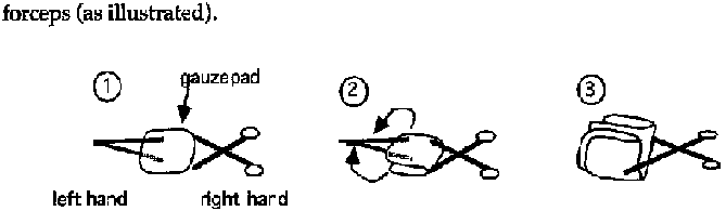
Figure
- Pour an antiseptic solution on the pad (infected wound, burns, abcess, ulcers: chlorhexidine-cetrimide; non infected surgical wound: polyvidone iodine; see table 25).
- Clean the periphery of the wound either with a circular movement, or from top to bottom. Change gauze pads as often as necessary.
- Clean the wound from top to bottom with a new tampon.
- Dry the periphery of the wound and then the wound itself with different gauze pads.
DRESSING A WOUND
- Apply one or several gauze pads to the wound.
- Apply strips of adhesive tape:
· perpendicularly to the axis of the limb or the
body;
· Leave the central part free to avoid maceration.
N.B.: When sterile disposable material is limited, sterile pads should be reserved for aseptic and surgical wounds.
Frequency of dressings
- Surgical wounds, or non infected sutures
· First day dressing should be well protected.
·
Further dressings, every 48 to 72 h (check the process of recovery).
- Infected wounds
· Dress every 24 h.
- Deep or large burns
· Dress on the first day, then leave until the 7th day (unless obvious infection).
- Phagedenic ulcers
· Dress every 24 h, with hospitalisation if possible.
Associated antibiotic treatment
As a rule, systemic antibiotic treatment should not be prescribed routinely.
- Deep and soiled wounds, to prevent gas gangrene procain-penicillin (IM): 4 or 5 IU per day x 5 days at least.
- Abcess
Antibiotic treatment is useless before incision.
- Burns
Only if they are infected.
- During conflicts or other disaster relief conditions, where access to health care and patient's follow-up are hazardous, the systematic use of PPF(or procain-penicillin) should be considered.
Wastes
All soiled disposable materials (gauze, coton, dressings, etc...) should be collected and bumed daily.
Choice and use of antiseptics and disinfectants
See table 25.
Abscess
A collection of pus in the soft tissues. An abscess cavity is
not accessible to antibiotics.
Treatment is thus surgical only.
Indications
Incision and drainage (I & D) should be performed once the abscess is "ripe" i.e. fluctuant upon gentle palpation.
Material
- Sterile scalpel blade and handle.
- Surgical gloves.
- Plain curved forceps (Kelly forceps).
- Sterile corrugated drain.
- Antiseptic solution e.g. chloramine solution or chlorhexidine-cetrimide solution (preparation: see table 25).
- 5 or 10 ml syringe.
Anesthesia
Anesthesia of an abscess by local infiltration with lidocaine is not very effective. Furthermore, the act of traversing wider areas of tissue with a needle may spread the infection further. Regional anesthesia is preferable where possible: e.g. ring block of a finger. Otherwise, the skin can be briefly numbed using ethyl chloride spray.
General anesthesia may be necessary for an abscess that is large or deep such as some breast abscesses, "injection" abscesses of the buttock, and pyomyositis: ketamine 1-2 mg/kg by slow IV or 5-10 mg/kg IM. The smaller IV dose acts more rapidly and for a shorter time than an IM dose and may thus be preferable.
Technique
- Scalpel: the correct way to hold a scalpel is between the thumb and forefinger with the handle resting against the palm (see Figure 7a). It should not be held as one holds a pen. The plane of the scalpel blade should be perpendicular to the plane of the skin.
- Incision: the free hand immobilizes the wall of the abscess between thumb and forefinger. Incise in the long axis of the abscess with a single stroke to breach the skin. The incision should be long enough to allow insertion of an exploring finger.
- Precautions: take care not to incise too deeply if the abscess overlies major blood vessels (the carotid, axillary, humeral, femoral and popliteal regions). After breaching the skin, blunt dissect down to the cavity using Kelly's forceps.

figure
· Explore the cavity with the forefinger, breaking any loculating adhesions and evacuating the pus (see Figure 7b).
· Abundant lavage of the cavity using a syringe filled with chloramine solution or chlorhexidine-cetrimide solution (preparation: see table 25).
· Insert a drain, if possible fixing it with a single suture at the edge of the incision. The drain is withdrawn progressively then removed altogether after 3 to 5 days (see Figure 7c).
BREAST ABSCESS
(see Figures 8a to 8d)
- The management of breast abscess is slightly different. Usually the abscess is superficial but deep ones, when they occur, are more difficult to diagnose and to treat.
- Early in the infection, before the infection loculates (mastitis), non-surgical measures should be applied:
· Antibiotics:
ampicillin(PO): 100 mg/kg/d x 5
days
or
chloramphenicol (PO): 75 mg/kg/d x 5 days.
· Anti-inflammatories:
indomethacin (PO): 75 mg/d
divided in 3 doses x 3 days
· Hot compresses, a constricting bandage to reduce lactation in the affected breast and expression of milk to avoid engorgement.
Material
- Same material as for other abscesses (see above).
Technique
- Incision:
· for superficial abscess: radial
· for abscess
near nipple: pert-alveolar
· for deep abcess: beneath the breast
- Gentle exploration with finger or Kelly forceps.
- Abundant
ravage with chloramine solution or chlorhexidine-cetrimide solution.
-
Insertion of drain.
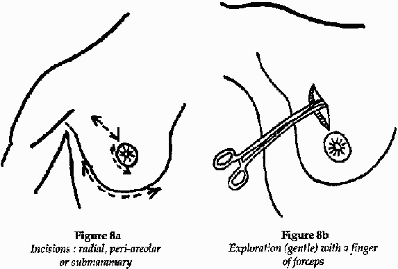
Figure 8a and 8b

Figure 8c and 8d
ABSCESS IN THE PAROTID REGION
There is a danger of sectioning the branches of the facial nerve. The incision should be over the caudal part of the abscess and parallel to the lower border of the maxilla (see Figure 9).
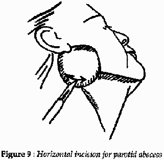
Figure
9
Pyomyositis
Infection and eventually abscess formation within muscde, most often due to Staphylococcus aureus.
At the start of infection, when the muscle is swollen, hot and painful, medical treatment may prevent abscess formation: immobilize, give antiinflammatory medication (indomethacin (PO): 75 mg/d divided in 3 doses x 5 days) and antibiotics (ampicillin (PO): Adult: 4 g/d in divided 3 doses;
Child:100 mg/kg/d divided in 3 doses x 7 days).
Indication
If the swelling becomes fluctuant conduct an exploratory puncture with a large-bore needle which will reveal thick pus.
Material
The same that for an abscess.
Anesthesia
Use ketamine (IM) if needed.
Technique for abscess drainage
- Generous skin incision, avoiding underlying neurovascular tracts, and incision of the fascia and muscle sheath, also with the scalpel (see Figure 10a).
- Blunt dissection with Kelly forceps down to the abcess cavity (see Figure 10b).
- Exploration with a finger to break adhesions and evacuate the pus (see Figure 10c).
- Abundant ravage with chloramine solution or chlorhexidine-cetrimide solution.
- Where possible, counter-incision of the skin near the edge of the abcess, cutting down onto a finger that is inserted deep in the cavity. The counter-incision should be anatomically posterior to the abscess to allow gravity drainage (assuming the patient will be supine during recovery). A strip of corrugated drain is threaded through the two incisions (see Figure 10d), fixed with a suture to the edge of the incision and withdrawn around the 5th day.
Note: Myositis of the right psoas muscle may present in a manner identical to that of acute appendicitis. Surgical evacuation is necessary.
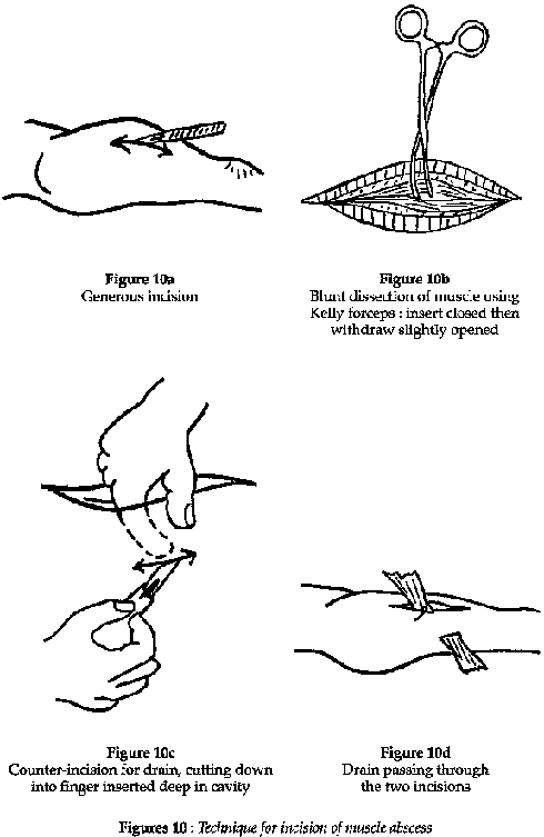
Figure
10
Burns
Thermal trauma to the skin, mucosa and deeper tissues. Burns are classified according to depth and extent.
Any burn that affects greater than 10 % of the body surface area is considered extensive and is thus serious because of fluid loss, cata-bolism, anemia and the risk of secondary infection. Burns are very comon in rural societies, particularly among children who fall onto or roll into cooking fires.
Clinical features
The extent of a burn is expressed as a percentage of total body surface area involved, easily estimated by the "rule of nines" (Table 26). The degree is a function of the depth to which tissue damage penetrates (Table 27).
A patient with extensive burns is likely to be in shock and requires appropriate resuscitation. Among children, the younger the patient the graver the danger presented by a burn of given extent and degree.
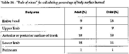
Table 26
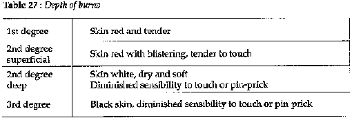
Table 27
Treatment
FIRST AID
- Immerse in cold water; this provides good analgesia and also arrests on-going trauma due to the heat retained in the tissues.
- Apply gentian violet.
- Do not cover.
RESUSCITATION
- Calculate the fluid requirements for the first 24 hours:
weight x 5% of surface burn x 2 = quantity of fluid required in rnls.
e.g.:
60 kg (wt) x 20 % (extent of burn)
60 x 20 x 2 = 2,400 ml
- 75 % of fluid should be given or ringer's lactate, the remainder as volume expanders or blood transfusion.
- During the first 24 hours, half the fluid requirements should be given in the first 8 hours.
FIRST DRESSING OF THE BURN
- Analgesia (pentazocine IM: 30 mg) and sedation if necessary (diazepam IM: 10 mg).
- Tetanus prophylaxis if available.
- Strict aseptic technique: drapes, gloves and instruments all sterile (Figure 11).
- Clean the burn with normal saline or ch/orhexidine-cetrimide solution (see table 25).
- Use a scalpel to debride blisters and non-viable tissue.
- Apply sterile vaseline gauze, then on top of that two layers of unfolded sterile gauze swabs. Do not use either antibiotic ointment or gauze impregnated with antibiotics or corticosteroids.
- Apply a bandage, not tightly. Do not wrap limbs, especially at the flexures as this will encourage contractures. Bandage each finger separately, never together.
- Immobilize limbs in the position of function.
- Alternatively: "open method": after wound cleaning leave the burn uncovered with the patient protected by a mosquito net.
SUBSEQUENT DRESSINGS
- Unless infection ensues, the first dressing should be left undisturbed for 5 to 7 days.
- Analgesia aseptic technique as for the first dressing.
- Remove any black eschars (which may hide purulent areas) and use scalpel to excise any necrotic tissue: skin, aponeurosis, muscle or tendon.
- Systemic antibiotics if obvious infection (not antibiotic
ointment):
PPF(or procain penicillin) (IM):
Adult: 4 MIU/d x 5 days at least
Child: 100,000 IU/kg/d x 5 days at least
- Same dressing as the first time. Again, this should not be removed for 5 to 7 days. Healing is signaled by granulation tissue: pink, mat and clean.
PATCH GRAFTING
(Figure 12)
- Skin grafting is necessary when the wound is slow to heal: often the case with deep second degree and third degree burns. Patch grafting is a simple technique and can also be used for treating tropical ulcers once the base is clean and granulating.
- Aseptic technique. Shave the donor area (usually anterior thigh or forearm) and prep with povidone iodine (see table 25). Infiltrate with lidocaine 1%.
- Lift up a patch of skin with fine toothed forceps and excise it with a scalpel. It should be full-thickness i.e. epidermis plus dermis. Take other patches from different parts of the donor site, leaving areas of intact skin between each excision.
- Spread each patch out on a sterile swab dampened with normal saline.
- Once a sufficient number of patches are excised, apply them carefully to the wound. Do not place them too close together: further healing will bridge the gaps and this allows a larger area to be grafted.
- Dress the donor and graft sites with sterile vaseline gauze, then layers of swabs and a non-compressive bandage.
- The graft will take within 7 days, during which time the dressing should not be removed and the patient should remain as immobile as possible.
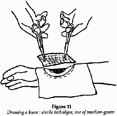
Figure 11
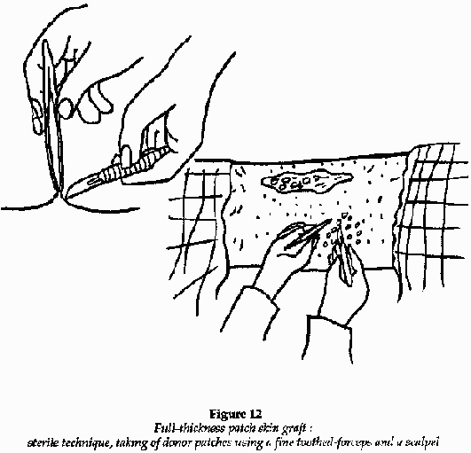
Figure
12
Wounds
General principles
This chapter concerns only wounds that can be treated at a dispensary level. For major trauma, refer to a surgical manual.
- Immediate ("primary") closure of wounds is desirable but not always practicable and in some circumstances it may be dangerous (risk of infection).
- Classically, it is said that a wound of greater than 6 hours should not be sutured. In isolated rural practice, however, patients often present late because of distances and this limit may be extended up to 24 hours, provided the patient can be observed during the following days for signs of infection.
- An infected wound should never be sutured.
- War wounds,
animal and human bites should not be sutured.
- Any break in the skin
overlying a fracture is an "open fracture".
- A wound that communicates with
a joint is an open joint wound.
- Always give antitetanus prophylaxis if
available.
The following are steps in the treatment of a wound: preparation, exploration, debridement, closure, drainage, and finally removal of sutures.
Preparation
WOUND TOILET
Shave if necessary, then clean the wound and its periphery with polyvidone iodine (see table 25).
MATERIAL
(Figures 13a to 13c and 14a to 14d)
- Sterile gloves and fenestrated drapes.
- Lidocaine, needle and syringe.
- Suture material.
- Suture set (sterilized box of instruments): needle holder, needles, scalpel blade and handle, one or two artery forceps, fine curved scissors with rounded ends, plain scissors for cutting sutures, retractors.
LOCAL ANESTHESIA
- Only necessary for large or deep wounds requiring more than 2
stitches.
- Lidocaine 1% without adrenaline.
- Infiltrate subcutaneously
via the wound edges.
Exploration
Once anesthetized, the wound can be explored and thoroughly cleaned of any debris.
Have a gloved assistant using retractors if necessary. Be careful to exclude the following:
- Foreign body.
- Underlying fracture.
- Involvement of
nerves, major blood vessels, tendons or joints.
- For scalp wounds:
underlying fracture (if serious may contain brain tissue).
Closure
- Use interrupted sutures (not continuous).
- Non-resorbable sutures such as silk for skin, resorbable thread (chromic catgut, Vicryl...) for subcutaneous tissues.
- Some suture material is already mounted on a needle by the manufacturer ("atraumatic needles").
- A curved needle is easier to manipulate.
- For skin use a "cutting" needle (triangular in cross-section); for subcutaneous tissues use a "round" needle (circular in cross-section).
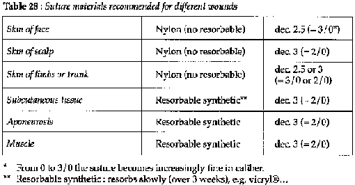
Table 28
Drainage
- Use a strip of corrugated rubber drain.
- Never use a drain for wounds of the face.
- Always insert a drain in wounds of the scalp and whenever a hematoma can be expected to form.
Removal of sutures
Face: day 5; other wounds: day 7 or 8.
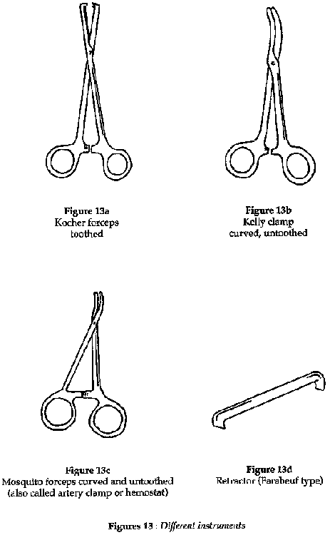
Figure 13
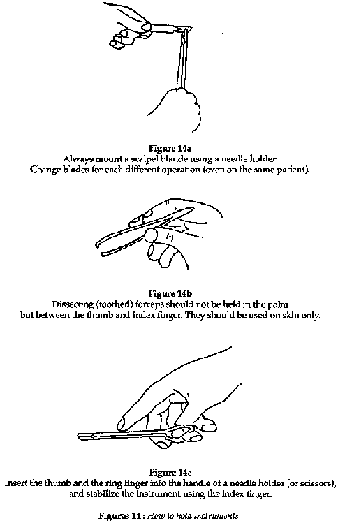
Figure 14
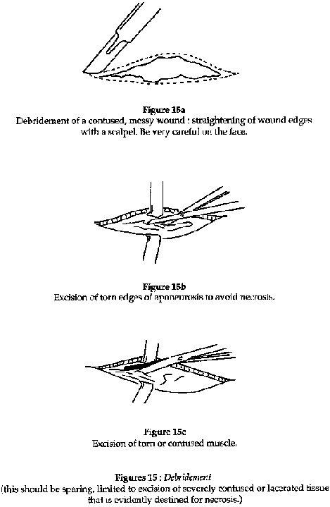
Figure 15

Figure 16
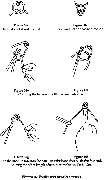
Figure 16
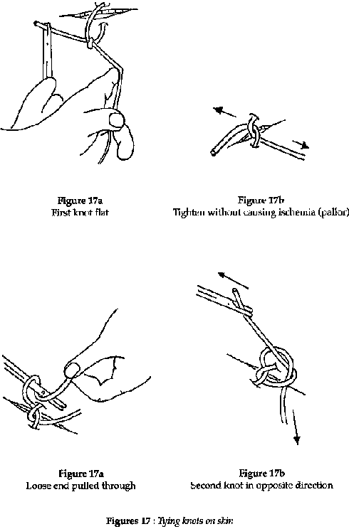
Figure 17
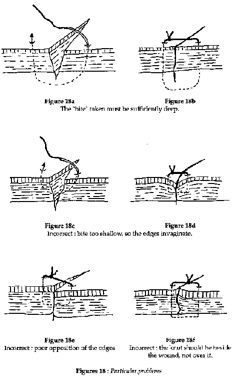
Figure 18
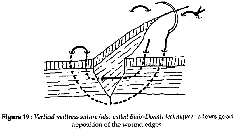
Figure 19
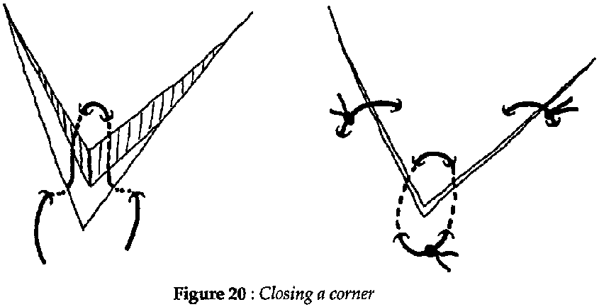
Figure 20
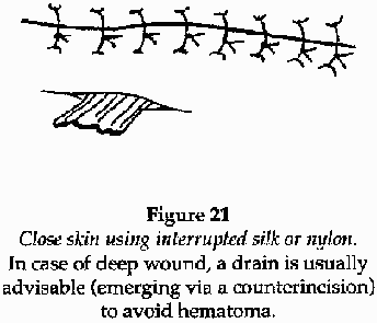
Figure 21

Figure
22
Bites and stings
Trauma caused by venomous animals; bites are inflicted by the mouth-parts (e.g. snakes, spiders), stings by the hindparts (e.g. bees, scorpions).
Treatment (hospital)
ENVENOMATION BY INSECTS, SCORPIONS AND SPIDERS
- Stings by bees, wasps...
Usually benign, but in susceptible
individuals may provoke either laryngeal edema or anaphylactic
shock:
adrenaline (epinephrine) (SC):
Adult: 1 mg
Child: 0.01 mg/kg
dexamethazone (IV): 4 mg stat. Repeat if required
plus a
perfusion of ringer´s lactate or volume expander.
- Spider bites and scorpion stings
Gravity depends upon the particular species, however the majority of such envenomations are either benign or else cause local tissue damage only. If a truly toxic species is thought to be responsible apply first aid and supportive measures as for snakebite (see below). Otherwise, therapy is limited to analgesia, local wound toilet and reassurance.
Clean and disinfect wound:
noramidopyrine (IM) (or any other
analgesic): 500 mg in 1 injection IM
If pain very severe:
pentazocine
(IM): 30 mg in injection IM
or lidocaine 1% (without adrenaline) infiltrated
around the wound gives good relief for very painful scorpion stings.
SNAKEBITE
It is most often not possible to identify the snake reponsible. In any case, the principles of management are the same: first aid and supportive therapy as indicated from close monitoring of the victim's clinical condition. Antivenenes are costly, difflcult to store, difficult to use, sometimes dangerous (anaphylaxis), and moreover of arguable efficacy.
- First aid: the "pressure-immobilization method". The object is to confine the venom to the site of the bite, thus allowing time for the body to metabolize it and for attendants to transport the victim to a health care facility. Venom diffuses mainly via the lymphatics, not via blood, tourniquets are thus of little use.
· Apply firm constant pressure to the site of the
bite.
· Apply a crepe bandage (or substitute) firmly to the entire
limb.
· Immobilize the limb with a splint.
· Immobilize the
patient.
- Supportive therapy: see table.
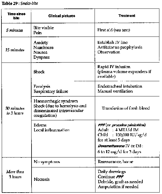
Table
29
|
| ||||||||||||||||||||||||||||||
Clinical Guidelines and Treatment Manual (MSF, 1993, 319 p.)
Appendix
Disinfection and Sterilization of medical equipment and supplies
- Sterilization = elimination of all micro-organisms (viruses, microscopic fungi, bacteria, both vegetative and spore forms).
- Disinfection = elimination of most micro-organisms present on a surface or object.
- Decontamination = disinfection of object soiled by infectious material (pus, blood, excrete...).
General rules
All equipment or supplies:
- coming into contact with sterile parts of the body (injection equipment, surgical instruments, some dressings, catheters...).
- used for perfusion.
should be sterilized and kept sterile
until utilization.
All reusable items, which do not correspond to the above definition, but which come into contact with mucus membranes, or get soiled with pus, blood, lymphatic or vaginal secretions, should be sterilized or subjected to a high level disinfection (effective among others against HIV and hepatitis B virus).
All soiled, non reusable equipment should be incinerated (warning: never recap needles after use = main cause of accidental needlestick).
To carry out proper sterilization is not always easy in the field conditions of isolated rural medical centers. It requires proper appliances (autoclave, hot air sterilizer), and an energy source.
In practice, one is often obliged to use alternative procedures which are not wholly satisfactory as they produce disinfection rather than sterilization (They are however compulsory if one cannot do better) (see following chapters).
Disinfection and sterilization of medical equipment is not enough to prevent iatrogenic infections (resulting from medical practice). It is obvious that basic hygienic and asepsis techniques ought to be applied: cleaning and disinfection of surfaces and premises, personal hygiene of the staff, aseptic handling of sterilized instruments...
Cleaning of reusable equipment
Soiled items and instruments should be carefully cleaned before being sterilized or subjected to a final disinfection.
The presence of organic matter could protect germs against the action of a disinfectant or sterilizing agent, or could react against it, rendering it ineffective.
INSTRUMENTS
Cleaning can be done either with water alone, with water and soap (or detergent), or with water and a compound of disinfectant/detergent.
Cleaning with a disinfectant chemical aims mainly to reduce the risks of contamination for the staff, but it does not eliminate them completely.
The staff in charge of instrument cleaning should be aware of the contamination risks (AIDS, hepatitis B), they should wear thick plastic or rubber gloves, and be careful when they handle sharp instruments.
After use and before cleaning, all instruments and items should be soaked in water to avoid deposits drying up. A disinfectant could be added for a first decontamination (chloramine 20 g/1, lysol 50 g/1).
Metallic instruments can be damaged if they are left in water too long (over several hours) or if the disinfectant concentration is too high.
Note
Needles and syringes for immunization should be soaked and cleaned with water alone, as traces of soap and disinfectant can inactivate vaccines.
After cleaning, instruments and items should be rinsed thoroughly with water and dried, then sterilized, boiled or disinfected (with a high level disinfectant) depending on their use and the local sterilizing facilities.
LINEN AND DRESSING
To decontaminate linen and dressings, one should wash them with an ordinary washing powder (ea. OMO) and boil them if possible (5 minutes).
If boiling is not possible, linen should be washed, rinsed and soaked for 30 minutes, in a 0.1 % chlorine solution (hypochlorite, bleach, chloramine), or 5 % lysol solution. It should then be rinsed abundantly and dried.
Theatre linen should be sterilized in an autoclave or ironed depending on local facilities.
Sterilization methods and alternatives
AUTOCLAVING
Sterilisation by steam under pressure in an autoclave.
Autoclaving is the most reliable sterilization method and the only one that allows effective sterilization of all medical equipment and supplies (especially linen and rubber). But relatively sophisticated appliances and energy source (electricity, kerosene or gas) are needed.
It is based on the same principle as a kitchen pressure cooker. Because water is heated in a closed container, temperatures above 100°C can be reached.
In the absence of air (air is purged at the beginning of sterilization), the temperature can be regulated by controlling the pressure.
According to the type of supply to be sterilized, sterilisation is carried out at 121°C (1 atmosphere over atmospheric pressure) or at 134°C (2 atm. over atmospheric pressure).

Table
Note:
- Do not forget to expell air (purge) while increasing the pressure (otherwise the temperature in the autoclave will not be sufficient).
- Drums or boxes holding objects to be sterilized must be open, never closed (unless fenestrated). The sliding windows in the special autoclave boxes should also be open during sterilization.
- Count the sterilizing time from the moment the required temperature or pressure is reached, not from the start of the heating phase.
DRY HEAT (IN HOT AIR STERILIZER OR OVEN - CALLED A POUPINEL IN FRENCH)
Sterilization by hot air (dry heat) at 160°C (320°F) for 2 hours or at 170°C (340°F) for 1 hour.
Reliable method provided it is carried out in a good electric appliance with working thermometer (an air circulation device is needed in large ovens).
This method is convenient for metal, heat resistant glass, and vaseline, but is not convenient for linen or gauze swabs. The oven method is quite simple but consumes more energy than an autoclave.
Ovens heated by charcoal fires or kerosene heaters are not reliable because they do not produce a sufficiently high temperature.
Time should be calculated from the moment the required temperature is reached (this is very important).
Notes
- Begin heating with the door open to expel any humidity (which could rust instruments).
- Do not exceed 170°C (could damage metallic instruments).
- It is better to place items in closed boxes. However, large boxes should be left halfopen to allow the material to more rapidly achieve the correct temperature.
BOILING
Boiling for 20 min (adding 5 minutes for 1000 altitude) provides high level disinfection, but not sterilization because it does not destroy bacterial spores (eg.: tetanus, gangrene).
Boiling is nevertheless essential when autoclaving or hot air sterilization are not possible. It is particulary useful for needles and syringes (it destroys HIV and hepatitis B virus).
After needles and syringes have been boiled, they should be kept dry and not left in the water (which can easily become recontaminated).
FLAMING
- In a flame: Effective if instruments are made red hot. This method should only be used in exceptional circumstances as it damages metal.
- With alcohol: Instruments are dipped in alcohol and set alight. This method is unreliable, expensive and in the long term damages instruments.
IRONING
Surgical drapes and gauzes can be ironed if an autoclave is either unavailable or too small to hold large operating drapes.
Iron on a table or bench covered with a sheet that has itself just been "sterilized" by ironing.
Dampen each item slightly with filtered boiled water.
The iron should be very hot and passed several times over each side of the linen/gauze.
However, if it is available, autodaving is always the preferred method.
IMMERSION IN "HIGH LEVEL" DISINFECTANTS
Immersion (of clean equipment) in the following disinfectant solutions destroys bacteria and virus including HIV and hepatitis B virus. The bacterial spores are generally not destroyed.
This process could be used as an alternative to sterilization when autoclaving or hot air sterilization are not possible.
Boiling however is always preferred. The effectiveness of chemical disinfection can always be impaired by dilution errors, by bad storage conditions, or by prolonged utilization of the same solution (solutions should be renewed at least once a day).
Chemical disinfection should never be recommended for syringes and needles.

Table
1. Hypochlorite solution (0.1 % or 1,000 ppm-1 ppm=1part per million=1mg/l- available chlorine) is prepared either from liquid bleach recently manufactured (< 3 months) or from calcium hypochlorite or from sodium dichloroisocyanurate (NaDCC, "Javel tablets", Javel solid, Stafilex, Actisan...), diluted according their respective available chlorine content.
Fresh liquid bleaches contain 3 to 15 % available chlorine (sometimes expressed in chlorometric degrees, 1° chlorom. = approx. 0.3 % available chlorine). Calcium hypochlorite contents from 30 to 70 % available chlorine. The NaDCC based tablets content generally 1.5 g available chlorine per tablet (1 tablet per litre = 1,500 ppm available chlorine).
NaDCC withstands heat much better than bleach and calcium hypochlorite.
2. As hypochlorite solutions are corrosive for metal, these solutions are convenient only for good quality stainless steel. The soaking should not exceed 1/2 hour and should be followed by thorough rinsing.
3. If instruments are used immediatly after soaking, it is not necessary to rinse the chloramine or the polyvidone iodine solution.
4. Ethanol and isopropylic alcohol (isopropanol) should be used at 70 % (70°) for the best effectiveness (more concentrated solutions are less effective). The prices, transportation and importation problems limit the use of these alcohols.
5. Immersion for several hours in aldehyde solutions, formaldehyde (formalin) and glutaraldehyde (Cidex), provides proper sterilization (destruction of all germs). These solutions however have many disadvantages: thorough rinsing compulsory (toxic residues), toxic vapours (formalin), high cost (glutaraldehyde).
Notes
- In order to obtain effective disinfection, eguipment must be cleaned before immersion in all these solutions...
- Aqueous solutions of cetrimide (Cetavlon), chlorhexidine (Hibitane), Savlon, HAC, Dettol and other common detergent and disinfectant solutions do not provide sufficient disinfection.
Soaking instruments in these solutions with the aim of "sterilization" should be avoided. This only provides an illusive feeling of safety and could in fact be a source of contamination.
STERILIZING GASES
- Ethylene oxide.
This method cannot be considered in field conditions because of its cost and of the special installation it requires (ethylene oxide is very toxic).
- Formol vapour (paraformaldehyde or trioxymethylene or "formol" tablets and Aldhylene)
Formol autoclaving also cannot be considered in the field. However formol vapour is often used for "makeshift" sterilization of instruments. The instruments are thoroughly cleaned and dried, then placed in a airtight container for at least 24 hours (minimum temperature of 20°C), either along with formol tablets 5 tablets for 1 litre container), or with formol alcoholic solution (Aldhylene) (1 ml for 1 litre container). Afterwards instruments are rinsed with sterile water. This is often impracticable, but it is absolutely compulsory if there is any visible deposit.
Users should be cautious during manipulation as vapors are toxic and highly irritative.
This method is not suitable for linen or gauze swabs as they absorb formaldehyde, which is toxic and necroses skin and mucus membranes.
This method is not totally reliable and has many disadvantages. It should be abandoned. If it is used an effective disinfection method against HIV (AIDS virus) (eg. boiling) should always be carried out before hand.
Equipment and metbods recommended
DISPENSARIES
Recommended equipment
- 1 small autoclave pressure cooker type (volume 15 to 20
litres)
- 1 powerful kerosene stove (or electric hot-plate)
- 1 metal mesh
basket
- Appropriate fenestrated containers (drums)
Recommended methods
- Instruments, syringes, glass, rubber, plastic, gauze swabs, small drapes: autoclave.
- Large drapes, gowns: wash with soap powder, boil if possible, then "sterilize" by ironing.
MOBILE TEAMS
Recommended equipment
If possible same equipment as for dispensaries.
Otherwise:
- 1 container for boiling
- Chloramine T or Polyvidone iodine
(Betadine)
Recommended methods
As for dispensaries if possible.
Otherwise:
- Metal instruments: boiling (best), otherwise sodium dichloroisocyanurate (NaDCC) or chloramine T or polyvidone iodine (exceptionally, after boiling and drying, instruments may be kept with formol tablet or Aldhylene until utilization)
- Needles, syringes: boiling
- Swabs: use disposable supplies
HOSPITALS WITH SURGICAL FACILITIES
Recommended equipment
Same equipment as for dispensaries and:
- 1 large autoclave (interior dimensions about 40 x 60 cm), operating with electricity, gas or kerosene according to local conditions.
- 2 mesh baskets
- Several fenestrated drums (number according to activity)
- Several fenestrated instrument boxes
If electric current is
available continuously for at least 3 hours per day:
- 1 electric hot air sterilizer
Recommended methods
- Metal instruments, glass: hot air sterilizer if good electric apparatus available, other-wise autoclave
- Swabs, linen (gowns, drapes): large autoclave
- Rubber, plastic items, syringes: small or large autoclave (at 121°)
Directions for use of an autolcave

Figure
1. Body of the autoclave
2. Mesh basket to contain packages to be sterilized
3. Metal base to support basket, drums above water
4. Drain tap
5. Lid, usually with a rubber seal and bolt-type catches
6. Tap or valve to allow purging of air during heating phase
7. Pressure valve: regulates the pressure by allowing excess vapour to escape
8. Safety valve
9. Pressure gauge
Note
In small autoclave, pressure cooker type, there is no purging tape, one uses the valve for purging.
The safety valve should not be manipulated during autoclaving (it will function only in case of excessive pressure rise).
Pressure gauge shows a pressure scale and sometimes a temperature scale. Pressure could be indicated in different manners.
One may consider that 1 bar = 1 kg/cm2 = 1 atmosphere = 15 psi
Temperature could be indicated in °C ou °F (135°C = 275°F; 121°C = 250°F).
OPERATION
1. Put the require quantity of water in the autoclave before each sterilization (dry heating could damage the autoclave): the level is usually marked or the quantity indicated by the manufacturer. If possible use distilled water or filtered rain water.
2. Place the objects to be sterilized in the mesh basket or onto the support, leaving enough room for vapour to circulate freely. The sliding "windows" on drums or containers must be open. Do not overload the autoclave.
3. Close the lid by tightening the bolds in diametrically opposite pairs (as the wheel nuts of a vehicle).
4. With the purging tap or valve open, begin to heat.
5. When a continuous jet of vapour is coming out of this tap/valve, close it.
6. Allow the pressure to rise to 0.5 atm, then open the purge tap/valve for 10 seconds to purge air, then close it.
7. Repeat this purge at about 0.7 atm, then again at about 0.9 atm.After this, al1 air should have been expelled from the autoclave and only steam will remain.
8. When desired operating pressure (and thus temperature) is obtained, sterilization begins. Start to time it then, not before.
The pressure valve regulates the pressure inside the autoclave allowing excess steam escape. There may be two interchangeable valves or positions to operate at either 1 or 2 atm.. If a lot of steam is being expelled, heat source should be lowered slightly.
9. After the required duration of sterilization, shut off the heat source.
10. Evacuate water and steam:
- For large autoclaves: through drain tap (to be connected outside).
- For pressure cooker type autoclaves: evacuate the steam by opening the purge valve. Once pressure drops to zero, open the lid, lift out the basket, pour out the water then replace the basket.
11. Allow to cool with the lid slightly open. Residual heat helps dry the sterilized items (the danger of contamination by ambient air is minimal).
12. Once items are dry, close the sliding windows on drums.
Note
If the autoclave is equiped with a drying system, follow the manufacturer's recommendations starting from paragraph 9.
PRESSURE OR TEMPERATURE AND DURATION REQUIREMENTS

Table
OPERATION VERIFICATION
- The stove should be powerfull enough to obtain a minimum rise of pressure of 1 atmosphere (1 bar or 1 kg/cm2 or 15 Psi).
- If possible, use sterilization autoclave tests, for example, 3M Autoclave Tape should turn black, brown is insufficient).
Warning, do not confuse test tape for hot air sterilizers with that for autoclaves. They are very similar but not interchangeable.
Place tests (eg. strip of tape) in the middle of the load into the boxes or drums to ensure that sterilization (temperature, steam, duration) is completed.
PACKAGING OF ITEMS FOR STERILIZATION
- Packaging of items: either
· without package if items are to be used
immediatly,
· fenestrated drums or boxes,
· heavy duty paper:
wrapping paper, kraft paper or news paper (2 layers),
· closely-woven
linen (2 layers),
· mixed (1 layer of paper, 1 layer of linen).
Paper plus linen is advisable if item is to be stored several weeks (because more resistant than paper alone and best barrier for germs than linen alone).
- Fenestrated containers should be equiped with a filter (a layer of heavy duty paper see above) accross the windows within the container or around the load to be sterilized so as to filter air during the drying phase after auto-craving. The paper should be checked and renewed regularly.
- If the autoclave is not equiped with a drying system, drying up of items inside boxes and drums is often unsastifactory. It is easier when the items are packed with paper or linen.
- Packed items should be placed vertically in the autoclave basket (not lying flat).
- Small packages and small drums are preferable to large ones.
- Needles and syringes: separate plunger and barrel of syringes and stick needles onto a gauze swab.
- Swabs and drapes should not be compressed inside boxes or drums.
Monthly epidemiological report
The goal of this report is to facilitate and standardize data collection for epidemiological surveys. It should record the monthly activities of the program and help in constructing the three-month and yearly reports. This form is a frame work for data collection, it should be adapted to the specific program.
Identification
|
Country: |
Month: |
|
Place or site: |
Year: |
Population
MONTHLY REPORT
|
Source: |
Total of previous month:
|
Arrivals: | |
+ |
-Departures: |
|
|
Births: | |
+ |
-Deaths: | |
| | | | |
Monthly report |
| |
+ Subtotal: |
|
- Subtotal: |
|
Average population= (total of previous months + monthly total)/2
AGE DISTRIBUTION
|
Source: | ||
|
Methodology of data collection: |
Survey |
Census |
|
Date of data collection: | ||
| |
Male |
Female |
0-4 years |
5-14 years |
15-44 years |
>=45 years |
Total |
|
% | | | | | | |
100% |
|
Number | | | | | | | |
Medical staff
The "title" (diploma, qualification) of each member of the medical staff should be indexed in the table below:
Table
Mortality
The data collection should be carried out by the administration in charge of the civilian status in order to obtain the most representative date (counting death that occured outside of health structures).
The personnel in charge of death records (political authorities, administrative, religious...) should be trained. This training consists of describing the most frequent pathologies and how to create a new file. One is only concerned with the primary cause of death.
|
Source of data collection: |
|
Table: |

Table
Morbidity
Record of new cases diagnosed.

Table
Rules for morbidity data collection
- The information is collected at the O.P.D. by physicians, nurses, medical auxiliaries; the medical staff will be supervised to make sure that definitions are respected.
- Only the new cases are recorded: patients consulting again for the same reason, in the same month, will be recorded at the index "reconsultation".
- The diagnosis is the one mentioned at the consultation (only one diagnosis per patient).
Definition of the table index
- Fever is defined as temperature > 38°C (axilla).
- Upper respiratory tract infections: any nose ear throat infection (N.E.T.) (sinusitis, cold, otitis, pharyngitis, laryngitis...).
- Lower respiratory tract infections: any infectious episode below the larynx (bronchitis, pneumonia, bronchiolitis...).
- Malaria: any fever (complicated or not), related to malaria (specify the definition: clinical or proven by microscopic examiniation).
- Measles: fever, + rhinopharyngitis, + conjunctivitis, + one of the two following signs:
· koplick's spots
· skin eruption
- Eye infection: unilateral or bilateral conjunctival inflammation or infection of any other part of the eye: conjunctivitis, trachoma, keratitis...
- Diarrhea: any episode with more than 3 watery stools per day.
· watery: is an estimation frequency of viral and choleriform diarrhea.
· bloody: estimates the frequency of entero-invasive diarrhea (bacillary and amoebic dysenteria).
- Cutaneous infection: any cutaneous infection due to a bacterial (impetigo, pyodermitis, abscess), viral (zone, herpes...), mycosal (ring worm...) or parasitic (scabies) infection.
- Sexually transmitted disease: genital infections, ulcerative or discharging (vaginitis, urethritis), apparently related to sexual contamination.
- Obstructive jaundice: yellow conjunctivitis, discolored stools, discolored urine and associated signs. It estimates the frequency of hepatitis.
- Urinary tract infections: burning on micturition associated with pollakiuria, whether there is fever, lumbar pain or not.
- Tuberculosis: the new cases begin their treatment during the month of diagnosis (bacteriological positive Ziehl's colouration for the pulmonary TB).
- Meningitis: any meningeal syndrome with fever, diagnosed by a physician.
- Trauma and burns: any consultation related to trauma (fight, fall, burn, wound...).
- Others: tetanus, poliomyelitis, diphtheria, whooping cough, typhus, leprosy, trypanosomiasis... adapt according to the situation. Each of these supplementary itemps will have to be defined by the team and the definition will be added to the one above.
List of essential drugs of WHO
(7th list, 1992)
1. Anaesthetics
1.1 GENERAL ANAESTHETICS AND OXYGEN
Diazepam (lb, 2) Ether,
anaesthetic (2) Halothane (2) Ketamine (2)
Nitrous oxide (2) Oxygen
Thiopental (2)
1.2 LOCAL ANAESTHETICS
Bupivacaine (2,9) Lidocaine
1.3 PREOPERATIVE MEDICATION
Atropine, Chloral hydrate,
Diazepam (lb) Morphine (la)
Promethazine
2 Analgesics, antipyretics, non-steroidal anti-inflammatory drugs and drugs used to beat gout
2.1 NON-OPIOIDS
Acetylsalicylic acid, Allopurinol (4)
Colchicine (7) Ibuprofen,
Indometacin, Paracetamol
2.2 OPIOID ANALGESICS
Codeine (1a)Morphine (l a)Pethidine (A)
(la, 4)
3. Antiallergics and drugs used in
anaphylaxis
Chlorphenamine, Dexamethasone,
Epinephrine.Hydrocortisone,
Prednisolone
4. Antidotes and other substances used in poisonings
4.1 GENERAL
Charcoal, activated Ipecacuanha
4.2 SPECIFIC
Atropine,Deferoxamine, Dimercaprol (2)
Methionine Methylthioninium chloride (methylene blue), Naloxone Penicillamine
(2) Potassium ferric hexacyanoferrate (II) 2H2O (Prussian blue), Sodium calcium
edetate (2) Sodium nitrite, Sodium thiosulfate
5. Antiepileptics
Carbamazepine, Diazepam (lb)Ethosuximide,
Phenobarbital (lb)Phenytoin, Valproic acid (7)
6. Anti-infective drugs
6.1 ANTHELMINTHICS
6.1.1 Intestinal anthelminthics
Levamisole (8) Mebendazole,
Niclosamide, Piperazine, Praziquantel, Pyrantel, Tiabendazole
6.1.2 Specific anthelminthics
Albendazole
6.1.3 Antifilarials
Diethylcarbamazine, Ivermectin, Suramin
sodium (2, 7)
6.1.4 Antischistosomals
Metrifonate, Oxamniquine,
Praziquantel
6.2 ANTIBACTERIALS
6.2.1 Penicillins
Amoxicillin (4) Ampicillie (4) Benzathine
Benzyl penicillin (5) Benzylpenicillin, Cloxacillin Phenoxymethyl penicillin,
Piperacillin, Procaine Benzylpenicillin
6.2.2 Other antibacterials
Chloramphenicol (7) Ciprofloxacin
(B) Clindamycin (B) Doxycycline (B) (5, 6) Erythromycin, Gentamicin (2, 4, 7)
Metronidazole, Nitrofurantoin (B) (4, 7) Spectinomycin (8) Sulfadimidine
(4)
Sulfamethoxazole + trimethoprim (4) Tetracycline, Trimethoprim (B)
6.2.3 Antileprosy drugs
Clofazimine, Dapsone, Rifampicin
6.2.4 Antituberculosis drugs
Ethambutol (4)
Isoniazid,Pyrazinamide, Rifampicin, Rifampicin + isoniazid, Streptomycin (4)
Thioacetazone + isorniazid (A) (7)
6.3 ANTIFUNGAL DRUGS
Amphotericin B(4) Flucytosine (B) (4,
8)Griseofulvin
6.3 ANTIFUNGAL DRUGS
Ketoconazole (2)Nystatin
6.4 ANTIPROTOZOAL DRUGS
6.4.1 Antiamoebic and antigiardiasis drugs Chloroquine (B) Diloxanide, Metronidazole
6.4.2 Antileishmaniasis drugs Meglumine antimoniate,Pentamidine (5)
6.4.3 Antimalarial drugs a) For curative treatment, Chloroquine,Mefloquine (B),Primaquine,Quinine, Tetracycline (B), Sulfadoxine + pyrimethamine (B) b) For prophylaxis, Chloroquine,Mefloquine (B),Proguanil
6.4.4 Antitrypanosomal drugs a) African trypanosomiasis,Eflornithine (C),Melarsoprol (5),Pentamidine (5),Suramin sodium b) American trypanosomiasis Benzonidazole (7),Nifurtimox (2, 8)
6.5 INSECT REPELLENTS
Diethyltoluamide
7. Antimigraine drugs
7.1 FOR TREATMENT OF ACUTE ATTACK
Acetylsalicylic
acid,Ergotamine (7),Paracetamol
7.2 FOR PROPHYLAXIS
Propanolol
8. Antineoplastic and immunosuppressant drugs
8.1 IMMUNOSUPPRESSANT DRUG
Azathioprine (2) Cidosporin (2)
8.2 CYTOTOXIC DRUGS
Bleomycin (2), Cisplatin (2),
Cydophosphamide (2), Cytarabine (2), Dacarbazine (2), Dactinomycin (2),
Doxorubici (2), Etoposide (2), Fluorouracil (2), Mercaptopurine (2),
Methotrexate (2), Procarbazine, Vinblastine (2), Vincristine (2)
8.3 HORMONES AND ANTIHORMONES
Dexamethasone,
Ethinylestradiol, PrednisoloneTamoxifen
9. Antiparkisonism drug.
Biperiden, Levodopa + Carbidopa (5,
6)
10. Drugs afecting the blood
10.1 ANTIANAEMIA DRUGS
Ferrous salt, Ferrous salt + Folic
acid,Folic acid (2),Hydroxocobalamin (2), Iron dextran (B) (5)
10.2 DRUGS AFFECTING COAGULATION
Desmopressin (8), Heparin,
Phytomenadione, Protamine sulfate, Warfarin (2, 6)
11. Blood products and plasma substitutes
11.1 PLASMA SUBSTITUTES
Dextran 70, Polygeline
11.2 PLASMA FRACTIONS FOR SPECIFIC USES
Albumin human (2,
8),Factor VIII concentrate(C) (2, 8) Factor IX complex concentrate (C) (2, 8)
12. Cardiovascular drugs
12.1 ANTIANGINAL DRUGS
Atenolol (B),Glyceryl
trinitrate,Isosorbide dinitrate,Nifedipine,Propranolol
12.2 ANTIDYSRHYTHMIC DRUGS
Atenolol
(B),Lidocaine,Procainamide (B),Propranolol,Quinidine (A),Verapamil (8)
12.3 ANTIHYPERTENSIVE DRUGS
Atenolol (B),Captopril
(B),Hydralazine,Hydrochlorothiazide,Methyldopa (B) (7),Nifedipine,Sodium
nitroprusside(C) (2, 8),Propranolol,Reserpine (A)
12.4 CARDIAC GLYCOSIDES
Digitoxin (B) (6) Digoxin (4)
12.5 DRUGS USED IN VASCULAR SHOCK
Dopamine
12.6 ANTITHROMBOTIC DRUGS
Acetylsalicylic acid, Streptokinase
(C)
13. Dermatological drugs
13.1 ANTIFUNGAL DRUGS (TOPICAL)
Benzoic acid + salicylic
acid,Miconazole, Nystatin, Sodium thiosulfate, Selenium sulfide (C)
13.2 ANTI-INFECTIVE DRUGS
Methylrosanilinium chloride
(gentian violet), Mupirocin, Neomycin
+ Q Bacitracin, Silver sulfadiazine
13.3 ANTI-INFLAMMATORY AND ANTIPRURITIC DRUGS
Betamethasone
(3) Calamine lotion, Hydrocortisone
13.4 ASTRINGENT DRUGS
Aluminium diacetate
13.5 KERATOPLASTIC AND KERATOLYTIC DRUGS
Salicylic
acid,Dithranol, Fluorouracil, Coal tar, Benzoyl peroxide, Podophyllum resin (7)
13.6 SCABICIDES AND PEDICULICIDES
Benzyl benzoate, Permethrin
13.7 ULTRAVIOLET-BLOCKING AGENTS
Benzophenones, sun
protection factor 15 (C) p-aminobenzoic acid, sun protection factor 15 (C), Zinc
oxide (C)
14. Diagnostic agents
14.1 OPHTHALMIC DRUGS
Fluorescein, Tropicamide
14.2 RADIOCONTRAST MEDIA
Amidotrizoate, Barium sulfate,
Iopanoic acid, Meglumine iotroxate (C), Propyliodone
15. Disinfectants and antiseptics
15.1 ANTISEPTICS
Chlorhexidine, Hydrogen peroxide, Iodine
15.2 DISINFECTANTS
Calcium hypochlorite, Glutaral
16. Diuretics
Amiloride (4, 7, 8), Furosemide,
Hydrochlorothiazide, Mannitol (C), Spironolactone (C)
17. Gastrointestinal drugs
17.1 ANTACIDS AND OTHER ANTIULCER DRUGS
Cimetidine, Aluminium
hydroxide, Magnesium hydroxide
17.2 ANTIEMETIC DRUGS
Metoclopramide, Promethazine
17.3 ANTIHAEMORRHOIDAL DRUGS
Local anaesthetic, astringent
and antiinflammatory drug
17.4 ANTI-INFLAMMATORY DRUGS
Hydrocortisone, Sulfasalazine
(2)
17.5 ANTISPASMODIC DRUGS
Atropine
17.6 CATHARTIC DRUGS
Senna
17.7 DRUGS USED IN DIARRHEA
17.7.1 Oral rehydration
Oral rehydration salts (for
glucose-electrolyte solution):
Sodium chloride 3.5 g/1, Potassium chloride
1.5 g/1, Trisodiurn citrate dihydrate 2.9 g/1, Glucose 20 g/1
17.7.2 Antidiarrheal (symptomatic) drugs
Codeine (la)
18. Hormones, other endocrine drugs and contraceptives
18.1 ADRENAL HORMONES AND SYNTHETIC
SUBSTITUTES
Dexamethasone, Fludrocortisone (C), Hydrocortisone, Prednisolone
18.2 ANDROGENS
Testosterone (C)
18.3 CONTRACEPTIVES
Depot medroxyprogesterone acetate (B) (7,
8), Ethinylestradiol + levonorgestrel, Ethinylestradiol + Norethisterone,
Norethisterone (B), Norethisterone enantate (B) (7, 8)
18.4 ESTROGENS
Ethinylestradiol
18.5 INSULINS AND OTHER ANTIDIABETIC AGENTS
Insulin injection
(soluble), Intermediate-acting insulin, Tolbutamide
18.6 OVULATION INDUCERS
Clomifene (C) (2, 8)
18.7 PROGESTOGENS
Norethisterone
18.8 THYROID HORMONES AND ANTITHYROID DRUGS
Levothyroxine,
Potassium iodide, Propylthiouracile
19. Immunologicals
19.1 DIAGNOSTIC AGENTS
Tuberculin, purified protein
derivative (PPD)
19.2 SERA AND IMMUNOGLOBULINS
Anti-D immunoglobulin (human),
Antiscorpion sera Antitetanus immuno globulin (human), Antivenom sera,
Diphtheria antitoxin Immunoglobulin human normal (2), Rabies immunoglobulin
19.3 VACCINES
19.3.1 For universal immunization
BCG vaccine
(dried)
Diphtheria-pertussistetanus vaccine, Diphtheria-tetanus vaccine,
Measles-mumps-rubella vaccine, Measles vaccine, Poliomyelitis
vaccine(inactivated), Poliomyelitis vaccine(live attenuated), Tetanus vaccine
19.3.2 For specific groups of individuals
Hepatitis B
vaccine, Influenza vaccine, Meningococcal vaccine, Rabies vaccine,Rubella
vaccine, Typhoid vaccine, Yellow fever vaccine
20. Muscle relaxants (peripherally acting) and cholinesterase
inhibitors
Gallamine (2), Neostigmine, Pyridostigmine (B) (2, 8),
Suxamethonium (2), Vecuronium bromide (C)
21. Ophthalmological preparations
21.1 ANTI-INFECTIVE AGENTS
Gentamicin, Idoxuridine, Silver
nitrate, Tetracycline
21.2 ANTI-INFLAMMATORY AGENTS
Prednisolone
21.3 LOCAL ANAESTHETICS
Tetracaine
21.4 MYOTICS AND ANTIGLAUCOMA DRUGS
Aeetazolamide,
Pilocarpine, Timolol
21.5 MYDRIATICS
Atropin Epinephrine (A)
22 Oxytocics and antioxytocics
22.1 OXYTOCICS
Ergometrine, Oxytocin
22.2 ANTIOXYTOCICS
Salbutamol (2)
23. Peritoneal dialysis solution
Intraperitoneal dialysis
solution (of appropriate composition)
24. Psychotberapeutic drugs
Amitriptyline, Chlorpromazine,
Diazepam (lb), Fluphenazine (5), Haloperidol, Lithium carbonate (2, 4)
25. Drugs acting on the respiratory tract
25.1 ANTIASTHMATIC DRUGS
Cromoglicic acid (B), Aminophylline
(2), Beclometasone, Ephedrine (A), Epinephrine, Salbutamol
25.2 ANTITUSSIVES
Codeine (la)
26. Solutions correcting water, electrolyte and acid-base disturbances
26.1 ORAL REHYDRATION
Oral rehydration salts (for
glucose-elecrolyte solution)
Potassium chloride
26.2 PARENTERAL
Compound solution of sodium lactate, Glucose,
Glucose with sodium chloride, Potassium chloride (2) Sodium chloride, Sodium
hydrogen carbonate
26.3 MISCELLANEOUS
Water for injection
27. Vitamins and minerals
Ascorbic acid (C), Calcium
gluconate (C) (2, 8), Ergocalciferol, Iodine, Nicotinamide, Pyridoxine, Retinol,
Riboflavin, Sodium fluoride (8), Thiamine
Many drugs included in the list are preceded by a square symbol to indicate that they represent an example of a therapeutic group and that various drugs could serve as alternatives. Numbers in parentheses following the drug names indicate:
(1) Drugs subject to international control under: a) the Single
Convention on Narcotic Drugs (1961), b) the Convention on Psychotropic
Substances (1971), or c) the Convention on Illicit
Traffic in Narcotic Drugs
and Psychotropic Substances (1988).
(2) Specific expertise, diagnostic precision or special equipment required for proper use.
(3) Greater potency or efficacy.
(4) In renal insufficiency, contraindicated or dosage adjustments necessary.
(5) To improve compliance.
(6) Special pharmacokinetic properties.
(7) Adverse effects diminish benefit/risk ratio.
(8) Limited indications or narrow spectrum of activity.
(9) For epidural anaesthesia.
Letters in parentheses after the drug names indicate the reasons for the inclusion of complementary drugs:
(A) When drugs in the main list cannot be made available.
(B) When drugs in the main list are known to be ineffective or inappropriate for a given individual.
(C) For use in rare disorders or in exceptional circumstances.
The New Emergency Health Kit - (WHO)
Lists of drugs and medical supplies for a population of 10,000 persons for approximately 3 months
Introduction
In recent years the various organizations and agencies of the United Nations system have been called upon to respond to an increasing number of large-scale emergencies and disasters, many of which pose a serious threat to health. Much of the assistance provided in such situations by donor agencies, governments, voluntary organizations and others is in the form of drugs and medical supplies. But the practical impact of this aid is often diminished because requests do not reflect the real needs or because these have not been adequately assessed. This can result in donations of unsorted, unsuitable and unintelligibly labelled drugs, or the provision of products which have passed their expiry date. Such problems are often compounded by delays in delivery and customs clearance.
The World Health Organization, which is the directing and coordinating authority for international health work within the United Nations system, took up the question of how emergency response could be facilitated. After several years of study, field testing and modifications, standard lists of essential drugs and medical supplies for use in an emergency were developed. The aim was to encourage the standardization of drugs and medical supplies used in an emergency to permit a swift and effective response with supplies that meet priority health needs. A further goal was to promote disaster preparedness since such standardization means that kits of essential items can be kept in readiness to meet urgent requirements.
The WHO Emergency Health Kit, which resulted from this work, was originally developed in collaboration with the Office of the United Nations High Commissioner for Refugees (UNHCR) and the London School of Hygiene and Tropical Medicine. It has now been revised in collaboration between the Action Programme on Essential Drugs (WHO, Geneva), the Emergency Preparedness and Response Unit (WHO, Geneva), the unit of Pharmaceuticals (WHO, Geneva), the Office of the United Nations High Commissioner for Refugees, UNICEF, Medecins Sans Frontieres, the League of Red Goss and Red Crescent Societies (Geneva), the Christian Medical Commission of the World Council of Churches and the International Committee of the Red Cross. A review of the experience of previous users of the kit, prepared by the London School of Hygiene and Tropical Medicine, as well as field experience of UNICEF and Medecins Sans Frontieres, were also considered during the revision. Major suppliers of the kit were consulted on the specifications of its contents.
The kit has now been adopted by many organizations and national authorities as a reliable, standardized, inexpensive, appropriate and quickly available source of the essential drugs and health equipment urgently needed in a disaster situation. Its contents are calculated to meet the needs of a population of 10,000 persons for three months. It has been renamed the: "New Emergency Health Kit" because of the number and diversity of United Nations agencies and other bodies which have adopted this list of drugs and medical supplies for their emergency operations and which participated in its revision.
This booklet provides background information on the development of the kit, a description of its contents, comments on the selection of items, treatment guidelines for prescribers and some useful checklists for suppliers and prescribers.
Chapter 1 (Essential drugs and supplies in emergency situations) is intended as a general introduction for health administrators and field officers.
Chapter 2 (Comments on the selection of drugs, medical supplies and equipment included in the kit) contains more technical details and is intended for prescribers.
Publication of this document was made possible by financial contributions received from the United Nations High Commissioner for Refugees, the Government of the Netherlands, the WHO Emergency Preparedness and Response Unit and the WHO Action Programme on Essential Drugs.
Chapter 1: Essential drugs and supli'es in Emergency situations
What is an Emergency ?
The term "emergency" is applied to various situations resulting from natural, political and economic disasters. The New Emergency Health Kit is not intended for the acute phase of epidemics, war, earthquake, floods, etc. but is designed to meet the needs of a population with disrupted medical facilities in the second phase of a natural or other disaster, or a displaced population without medical facilities. It has also been used in countries with acute shortages of drugs due to economic reasons.
It must be emphasized that, although supplying drugs and medical supplies in the standard kits is convenient in the second phase of an emergency, specific local requirements need to be assessed as soon as possible and further supplies must be ordered accordingly.
Qunantification of drug requirements
Morbidity patterns (the relative frequency of different illnesses) may vary considerably between emergencies. For example, in emergencies where malnutrition is common morbidity rates may be very high. For this reason an estimation of drug requirements from a distance can only be approximate, although certain predictions can be made based on past experience. For the present kit estimates have been based on the average morbidity patterns and the use of standard treatment guidelines. The quantities of drugs supplied will therefore only be adequate if prescribers follow these guidelines (given in Annexes 1-3).
Contents of the kit
The New Emergency Health Kit consists of two different sets of drugs and medical supplies, named a BASIC UNIT and a SUPPLEMENTARY UNIT (The previous version of three lists: A-basic drugs; B-supplementary drugs; C-medical supplies and requirement for basic supplementary lists). To facilitate distribution to smaller health facilities on site, the quantities of drugs and medical supplies in the basic unit have been divided into ten identical units for 1,000 persons each.

Table
The BASIC UNIT contains drugs, medical supplies and some essential equipment for primary health care workers with limited training. It contains twelve drugs, none of which are injectable. Simple treatment guidelines, based on symptoms, have been developed to help the training of personnel in the proper use of the drugs. Copies of these treatment guidelines, an example of which is printed in Annexes 1-3, should be be included in each unit. Additional copies can be obtained from the Action Programme on Essential Drugs, WHO, Geneva, and from UNICEF Copenhagen (see Annex 7 for addresses).
The SUPPLEMENTARY UNIT contains drugs and medical supplies for a population of 10,000 and is to be used only by professional health workers or physicians. It does not contain any drugs or supplies from the basic units and can therefore only be used when these are available as well.
The selection and quantification of drugs for the basic and supplementary units have been based on recommendations for standard treatment regimens from technical units within WHO. A manual describing the standard treatment regimens for target diseases, developed in collaboration between Medecins sans Frontieres and WHO, is available from Medecins sans Frontieres at cost price and is to be included in each supplementary unit.
To facilitate identification in an emergency, one green sticker (international color code for medical items) should be placed on each parcel. The word "BASIC" should be printed on stickers for basic units.
Referral system
Health services can be decentralized by the use of basic health care clinics (the most peripheral level of health care) providing simple treatment using the basic units. Such a decentralization will: 1) increase the access of the population to curative care; and 2) avoid overcrowding of referral facilities by solving all common health problems at the most peripheral level. Basic treatment protocols have been drawn up to allow these health workers to take the right decision on treatment or referral, according to the symptoms (see Annexes 1-3).
The first referral level should be staffed by professional health workers, usually medical assistants or doctors, who will use drugs, supplies and equipment from both the basic and the supplementary units. It should be stressed here that the basic and supplementary units have not been intended to enable these health workers to treat rare diseases or major surgical cases. For such patients a second level of referral is needed, usually a district or general hospital. Such facilities are normally part of the national health system and referral procedures are arranged with the local health authorities.
Procurement of the kit
The New Emergency Health Kit can be provided from a number of major pharmaceutical suppliers, some of which will have a permanent stock of kits ready for shipment within 48 hours. It may however be desirable to secure procurement at the regional level to reduce the cost of shipping. The procuring agency should ensure that manufacturers comply with the guidelines for quality, packaging and labelling of drugs (see Annexe 6).
It is important to note that many drugs in the kit can be considered as examples of a therapeutic group, and that other drugs can often serve as alternatives. This should be taken into consideration when drugs are selected at the national level, since the choice of drugs may then be influenced by whether equivalent products are immediately available from local sources, and their comparative cost and quality. National authorities may wish to stockpile the same or equivalent drugs and supplies as part of their emergency preparedness programme. The kit can also serve as a useful baseline supply list of essential drugs for primary health care.
Donor guidelines
Whatever the source of drugs, it is very important that:
- No drugs should be sent from a donor country without a specific request, or without prior clearance by the receiving country;
- No drugs should be sent that are not on the List of Essential Drugs of the receiving country, or, if such a national list is not available, on the WHO Model List of Essential Drugs;
- No drugs should arrive with a future life (before expiry date) of less than one year;
- Labelling of the drugs should be in the appropriate language(s) and should at least contain the generic name, strength, name of manufacturer and expiry date (see Annexe 6);
- Labelling on the outside package should contain the same information, plus the total quantity of drugs in the package.
Immunization in emergency
Experience in past emergencies involving displacements of populations has shown measles to be one of the major causes of death among younger children. The disease spreads rapidly in overcrowded conditions, and serious respiratory tract infections are frequent, particularly in malnourished children. An adequate supply of essential drugs may reduce the mortality rate, but measles can be prevented by immunization. A measles immunization programme should therefore be given high priority in the early phase of an emergency. The WHO Expanded Programme on Immunization (EPI), UNICEF, the Office of the High Commissioner for Refugees (UNHCR) and OXFAM have collaborated in the development of the Emergency Immunization Kit, which may be used to set up an emergency immunization programme against measles. This kit contains cold chain and injection equipment for 5,000 immunizations and may be ordered from OXFAM. Vaccines are not included.
Post emergency needs
After the acute phase of an emergency is over and basic health needs have been covered by the basic and supplementary units, specific needs for further supplies should be assessed as soon as possible. In most cases this will necessitate a quick description and, if possible, quantification of the morbidity profile. It should characterise the most common diseases and should identify the exposed and high risk groups in the population (e.g. children below 5 years of age and pregnant women). These high risk groups should be the first target of the continuing health care programme. Any other factors that may influence requirements should also be taken into account, ea. the demographic pattern of the community, the physical condition of the individuals, seasonal variations of morbidity and mortality, the impact of improved public health measures, the local availability of drugs and other supplies, drug resistance, usual medical practice in the country, capabilities of the health workers and the effectiveness of the referral system.
Much time and money may be saved by adapting re-order forms to the specific needs of the situation and by standardizing re-order procedures for all locations and health teams, regardless of whether supplies are available locally or must be ordered from abroad.
Chapter 2: Comments on the selection of drugs, medical supplies and equipment included in the kit
The composition of the New Emergency Health Kit is based on epidemiological data, population profiles, disease patterns and certain assumptions bome out by emergency experience. These assumptions are:
- The most peripheral level of the health care system will be staffed by health workers with only limited medical training, who will treat symptoms rather than diagnosed diseases and who will refer to the next level those patients who need more specialized treatment.
- Half of the population is 0-14 years of age.
- The average number of patients presenting themselves with the more common symptoms or diseases can be predicted.
- Standardized schedules will be used to treat these symptoms or diseases.
- The rate of referral from the basic to the next level is 10 %.
- The first referral level of health care is staffed by experienced medical assistants or medical doctors, with no or very limited facilities for inpatient care.
- If both the basic and first referral health care facilities are within reasonable reach of the target population, every individual will, on average, visit such facilities four times per year for advice or treatment. As a consequence the supplies in the kit, which are sufficient for approximately 10,000 outpatient consultations, will serve a population of 10,000 people for a period of approximately three months.
Selection of the drugs
Injectable drugs
There are no injectable drugs in the basic unit. Basic health workers with little training have usually not been taught to prescribe injections, neither are they trained to administer them. Moreover, the most common diseases in their uncomplicated form do not generally require an injectable drug. Any patient who needs an injection must be referred to the first referral level.
Antibiotics
Infectious bacterial diseases are common at all levels of health care, including the most peripheral, and basic health workers should therefore have the possiblity to prescribe an antibiotic. However, many basic health workers have not been trained to prescribe antibiotics in a rational way. Cotrimoxazole is the only antibiotic included in the basic unit, and this will enable the health worker to concentrate on taking the right decision between prescribing an antibiotic or not, rather than on the choice between several antibiotics. Cotrimoxazole has been selected because it is active against the most common bacteria found in the field, especially S. pneumoniae and H. influenzae for acute respiratory infections. It is also stable under tropical conditions, needs to be taken only twice daily and its side-effects (exfoliative dermatitis or bone marrow depression) are uncommon. In addition to this it is less expensive than other antibiotics. The risk of increasing bacterial resistance must be reduced by rational prescribing practice.
Drugs not included in the kit
The kit includes neither the common vaccines nor any drugs against communicable diseases such as tuberculosis or leprosy. The vaccines needed and any plans for an expanded programme on immunization should be discussed with the national authorities as soon as possible; the same applies for programmes to combat communicable diseases. In general no special programme should be initiated unless there is sufficient guarantee for its continuation over a longer period.
In addition, drugs in the kit do not cover some specific health problems occurring in certain geographical areas, e.g. specific resistant malaria strains.
Selection of renewable supplies
Syringes and needles
Considering the risk of direct contamination with hepatitis and AIDS during handling, needles are dangerous items. The health risk for the staff should be limited by the following means:
· Limiting the number of injections;
· Using
disposable needles only;
· Strictly following the destruction procedures
for disposable material.
It is less dangerous to handle syringes than needles. For this reason a system with resterilisable nylon syringes and disposable needles has been chosen for the supplementary unit. However, in the very first stage, when sterilization procedures are not yet established, some provision will be necessary for giving injections by means of fully disposable materials. A small number of disposable syringes are therefore provided in the supplementary unit and their destruction should be supervised by the person in charge.
Gloves
Disposable protective gloves are provided in the basic unit to protect health workers against possible infection during dressings or handling of infected materials. In any case a dressing should be applied or changed with the instruments provided in the kit. Surgical gloves, which should be resterilizable, are supplied in the supplementary unit. They are to be used for deliveries, sutures and minor surgery, all under medical supervision.
Selection of equipment
Resuscitation / Surgical instruments
The kit has been designed for general medicine under primitive conditions, and for that reason no equipment for resuscitation or major surgery has been included. In situations of war, earthquakes or epidemics, specialised teams with medical equipment and supplies will be required.
Sterilization
A complete sterilization set is provided in the kit. The basic units contain two small drums each for sterile dressing materials. Two drums are included to enable the alternate sterilization of one at the first referral level while the other is being used in the peripheral facility. The supplementary unit contains a kerosene stove and two pressure sterilizers, a small one for sterilizing 2 ml and 5 ml syringes, and a larger one for the small drums with dressing materials and the instrument sets.
Dilution and storage of liquids
The kit contains several plastic bottles and a few large disposable syringes which are needed to dilute and store liquids (e.g. benzyl benzoate, chlorhexidine and gentian violet solution).
Water supply
The kit contains several items to help provide for clean water at the health facility. Each basic unit contains a 20 litre foldable jerrycan and a plastic bucket. The supplementary unit contains a water filter with candles and 2.5 kg of chloramine powder to chlorinate the water.
Chapter 3: Composition of the New Emergency Health Kit
The New Emergency Health Kit consists of ten basic units and one supplementary unit.
10 basic units (for basic health workers) for a population of 10,000 persons for 3 months (1 basic unit for 1,000 persons for 3 months). The unit contains drugs, renewable supplies and basic equipment packed in one carton.
1 supplementary unit (for physicians and senior health workers), for a population of 10,000 people for 3 months. One supplementary unit contains:
- drugs (approximately 130 kg)
- essential infusions
(approximately 180 kg)
- renewable supplies (approximately 60 kg)
-
equipment (approximately 40 kg)
NB: The supplementary unit does not contain any drugs and medical supplies from the basic unit. To be operational, the supplementary unit should be used together with ten basic units.

Table
Basic unit (for 1,000 persons for 3 months)
Drugs
Acetylsalicylic acid, tab 300 mg.............................................................tab 3,000
Aluminium hydroxyde, tab 500 mg..........................................................tab 1,000
1) Benzyl benzoate, lotion 25 %...........................................................bottle 1 litre 1
2) Chlorhexidine (5%).........................................................................bottle 1 litre 1
Chloroquine, tab 150 mg base................................................................tab 2,000
Ferrous Sulfate + Folic Acid, tab 200 + 0.25 mg......................................tab 2,000
Gentian Violet, powder ..........................................................................25 g 4
Mebendazole, tab 100 mg ......................................................................tab 500
ORS (Oral Rehydration Salts) .............................................................. sachet for 1 litre 200
Paracetamol, tab 100 mg.......................................................................tab 1,000
Sulfamethoxazole + Trimetoprim, tab 400 + 80mg (cotrimoxazole) ............... tab 2,000
Tetracycline eye ointment 1 %............................................................... tube 5 g 50
Renewable supplies
Absorbent cotton
wool
................................................................ kg 1
Adhesive tape 2.5 cm x 5
m.......................................................... roll 30 Bar of soap
(100-200 g)............................................................... bar
10 Elastic bandage (crepe) 7.5 cm x 10
m
...............................................unit 20Gauze bandage 7.5 cm
x 10 m,.........................................................roll 100Gauze
compress 10 x 10 cm, 12 ply, nonsterile.......................................
unit 500Ballpen, blue or
black..............................................................
.unit
10Exercise book
A4
...................................................................
.unit
43) Health card + plastic
sachet......................................................unit 500Small
plastic bag for
drugs..........................................................unit
2,000Notepad
A6.................................................................
..........unit
10Thermometer (oral/rectal) Celsius
/
Fahrenheit...................................... unit 6Protective glove,
nonsterile, disposable............................................ unit
1004)
Treatment guidelines for basic
list.............................................. unit 2
Equipment
Nail brush, plastic,
autoclavable................................................... unit 2Bucket,
plastic, approx. 20
litres...................................................unit 1Gallipot,
stainless steel, 100 ml....................................................unit
1
1) According to WHO recommendations Benzyl henzoate solution 25 % concentration is being supplied. The use of 90 % concentration is not recommended.
2) Chlorhexidine 20 % needs distilled water for dilution, otherwise precipitation may occur. 5 % solution is WHO standard. Alternatives include the combination of chlorhexidine 1.5 % and cetrimide 15 %.
3) For a sample health card, see Annex 4.
4) For sample treatment guidelines, see Annexes 1, 2 and
3.
Kidney dish, stainless steel, approx. 26 x 14
cm.................................... unit 11) Dressing set (3 instruments +
box)............................................... unit 2Dressing tray,
stainless steel, approx. 30 x 15 x 3 cm.............................. unit 1Drum
for compresses approx. 15 cm H, 0 14 cm........................................
unit 2 Foldable jerrycan, 20
litres........................................................ unit 1Forceps
Kocher, no teeth, 12-14 cm..................................................
unit 2Plastic bottle, 1
litre
.............................................................unit
3Syringe Luer, disposable, 10
ml..................................................... unit 1Plastic bottle,
125 ml.............................................................. unit
1Scissors straight/blunt, 12-14
cm....................................................unit 2
1) Dressing set (3 instruments + box):
· 1 stainless steel box approx. 17 x 7 x 3 cm
· 1
pair surgical scissors, sharp/blunt, 12-14 cm
· 1 Kocher forceps, no
teeth, straight, 12-14 cm
· 1 dissecting forceps, no teeth, 12-14 cm
Supplementary unit (for 10,000 persons for 3 months)
Drugs
Anaesthesics
Ketamine, inj. 50
mg/ml........................................................10 ml/vial 251)
Lidocaine.
inj.
1%..........................................................20 ml/vial
50
Analgesics
2) Pentazocine, inj. 30
mg/ml............................................. 1 ml / ampoule 503)
Probenecid, tab 500
mg
............................................................tab 500Recall
from basic unit:Acetyl salicyclic acid, tab 300
mg.......................................... (10 x 3,000) 30,000Paracetamol, tab
100 mg
................................................... (10 x 1,000J)
10,000
Anti-allergics
Dexamethasone, inj. 4
mg/ml
...................................................1 ml / amp
50Prednisolone, tab 5
mg............................................................... tab
100Epinephrine (adrenaline), see "respiratory
tract"
Anti-epileptics
Diazepam, inj. 5
mg/ml.................................................... 2 ml / ampoule
200Phenobarbital, tab 50
mg
.............................................................tab 1,000
Anti-infective drugs
4) Ampicillin, tab 250
mg
........................................................... tab 2,0004)
Ampicillin, inj. 500 mg
/vial
................................................... vial 200Benzathine
benzylpenicillin, inj. 2.4 MIU / vial.................................... vial
50Chloramphenicol, caps 250
mg........................................................ caps
2,000Chloramphenicol, inj. 1 g /
vial.................................................... vial 500Metronidazole,
tab 250 mg............................................................ tab
2,0005) Nystatin, non-coated
tablet
..........................................100,000 IU / tab
2,000Phenoxymethylpenicillin, tab 250
mg
..................................................tab 4,0006) Procain
benzylpenicillin, inj. 3-4 MU / vial
....................................
vial 1,000
1) 20 ml vials are preferred, although 50 ml vials may be used as an alternative.
2) Because of narcotic drugs regulation, pentazocine has been chosen as an alternative to morphine or pethidine.
3) To be used with penicillin in the treatment of gonorrhoea.
4) Ampicillin tablets and injections to be used only in neonates and pregnant women.
5) For the treatment of oral candidiasis.
6) The combination of procaine benzylpenicillin 3 MU and benzylpenicillin 1 MU (procaine penicillin fortified) is used in many countries and may be included as an alternative.
1) Quinine, inj. 300
mg/ml
................................................... 2 ml / amp
100Quinine sulfate, tab 300
mg
..........................................................tab 3
,0002)
Sulfadoxine + pyrimethamine, tab 500 mg + 25
mg
.................................. tab 303) Tetracycline, caps or tab 250
mg
..........................................caps or tab 2,000
Recall from
basic unit
Mebendazole, tab 100
mg
...................................................... (10 x 500)
5,000Cotrimoxazole, tab 400 + 80
mg
..............................................(10 x 2,000)
20,000Chloroquine, tab 150
mg..................................................... (10 x
2,000) 20
Blood, drugs affecting the
Folic acid, tab 1 mg........................................................... 5,000
Recall from basic unit:
Ferrous sulfate + Folic acid, tab 200
+ 0.25 mg............................. (10 x 2,000) 20,000
Cardiovascular drugs
4) Methyldopa,
tab
250................................................................ tab
500Hydralazine, inj.:20
mg/ml
..................................................1 ml / amp 20
Dermatological
5) Polyvidone iodine 10 %, sol., 500
ml
...........................................bottle 4
Zinc oxyde 10 %
ointement..........................................................
.....kg
2Benzoic acid 6 % + salicylic acid 3 %
ointment........................................ kg 1
Recall from basic
unit:
Tetracycline eye ointment, 1
%
.................................................(10 x 50) 500Gentian
violet, powder 25 g..................................................... (10 x
4) 40Benzyl benzoate, lotion 25 %,
litre............................................. (10 x 1)
10
Diuretics
Furosemide, inj. 10
mg/ml..................................................... 2 ml / amp
20Furosemide, tab 40
mg
............................................................... tab
200
Gastro-intestinal drugs
Promethazine, tab 25
mg
..............................................................tab
500Promethazine, inj. 25
mg/ml................................................... 2 ml / amp 50.Atropine,
inj. 1 mg/ml
........................................................1 ml /
amp 50.
Recall from basic unit:
Aluminium hydroxyde, tab 500
mg...............................................(10 x 1,000)
10,000
1) For the treatment of cerebral and resistant malaria
cases.
Intravenous injection of quinine must always be diluted in 500 ml
glucose 5 %.
2) For the treatment of resistant malaria strains (check national protocols).
3) For the treatment of cholera and chlamydia infections.
4) For the treatment of hypertension in pregnancy.
5) Polyvidone iodine has been chosen because the use of iodine tincture in hot climates may result in toxic concentrations of iodine by partial evaporation of the alcohol.
Oxtocics
Ergometrine maleate, inj. 0.2
mg/ml..............................................1
ml/amp
200
Psychotherapeutic drugs
Chlorpromazine, inj. 25
mg/ml................................................. 2 ml / amp 20.
Respiratory tract, drugs acting on
Aminophylline,
tab
100mg.............................................................. tab
1,000Aminophylline, inj. 25
mg/ml
.................................................10 ml / amp
50Epinephrine (adrenaline), inj. 1
mg/ml
....................................... 1 ml / amp 50
Solutions
correcting water, electrolyte and acid-base disturbances(because of the weight,
the quantity of infusion included in the kit is minimal. Look for local supply,
once in the field.)
Compound solution of sodium lactate (Ringer's
Lactate),inj. sol., with giving set and
needle....................................... 500 ml / bag 200Glucose, inj. sol.
5 %, with giving set and needle...........................500 ml / bag 100(for
dilution of quinine/injection)Glucose, inj. sol.
50
%.................................................... 50 ml / vial 20Water
for injection
................................................ 10 ml /
plastic vial 2,000
Recall from basic unit:
ORS (Oral Rehydration
Salts)
................................................. (10 x 200)
2,000
Vitamins
Retinol (Vitamin A), caps
200,000
IU................................................ caps 4,000Ascorbic
acid, tab 250 mg
...........................................................
tab 4,000
Renewable supplies
Scalp vein infusion set, disposable, 25G
(�0.5 mm).................................. unit 300Scalp vein infusion set,
disposable, 21G (�0.8 mm).................................. unit 100IV placement
canula, disposable, 18G (�1.7 mm)...................................... unit
15IV placement canula, disposable, 22G (�0.9
mm)...................................... unit 15Needle Luer IV, disposable, 19G
(�1.1 mm x 38 mm)................................... unit 1,000Needle Luer IM,
disposable, 21G (�0.8 mm x 40 mm)................................... unit
2,000Needle Luer SC, disposable, 25G (�0.5 mm x 16
mm)................................... unit 100Spinal needle, disposable, 20G
(64 mm - �0.9 mm)
....................................unit 30Spinal needle,
disposable, 23G (64 mm - �0.7 mm).................................... unit
30Syringe Luer resterilisable, nylon, 2
ml............................................ unit 20Syringe Luer
resterilisable, nylon, 5 ml............................................ unit
100Syringe Luer resterilisable, nylon, 10
ml........................................... unit 40Syringe Luer, disposable, 2
ml
..................................................... unit 400Syringe
Luer, disposable, 5 ml
.....................................................
unit 500Syringe Luer, disposable, 10
ml
.................................................... unit 200Syringe conic
connector (for feeding), 60 ml
........................................unit
20Feeding tube, CH5 (premature baby),
disposable...................................... unit 10Feeding tube, CH8,
disposable....................................................... unit 50Feeding
tube, CH16, disposable......................................................
unit 10Urinary catheter (Foley), n°12,
disposable.......................................... unit 10Urinary catheter
(Foley), n°14, disposable.......................................... unit
5Urinary catheter (Foley), n°18,
disposable
..........................................unit 5Surgical gloves
sterile and resterilisable n°6.5.................................... pair
50Surgical gloves sterile and resterilisable
n°7.5
....................................pair 150Surgical gloves
sterile and resterilisable
n°8.5
....................................pair 50
Recall from basic
unit:
Protective glove, non sterile,
disposable............................... (100 units x 10)
1,000
Sterilization test tape (for
autoclave).............................................. rol 2Chloramine, tabs
or powder............................................................ kg
2,5Thermometer (oral/rectal) dual Celsius
/
Fahrenheit................................ unit 10Spare bulb for
otoscope...........................................................
. unit
2Batteries R6 alkaline AA size (for
otoscope)........................................ unit 6
Recall from basic
unit:Thermometer (oral/rectal)
celsius
/fahrenheit............................. (6 units x 10) 60Ballpen,
blue or black................................................... (10 units x 10)
100Exercise book A4
.........................................................
(4 units x 10) 40Health card + plastic
sachet
........................................... (500 units x
10)
5,000Small plastic baa for
drugs
.......................................... (2,000 units x
10)
20,000Notepad
A6
...............................................................(10 units x
10) 100
Urine collecting bag with valve, 2000
ml............................................ unit 10Finger stall 2 fingers,
disposable.................................................. unit 300
Suture, synthetic absorbable, braided, size DEC.2 (000)
withcutting needle curved 3/8, 20 mm
triangular......................................... unit 24
Suture, synthetic
absorbable, braided, size DEC.3 (00) withcutting needle curved 3/8, 30 mm
triangular......................................... unit 36Surgical blade
(surgical knives) n°22 for handle
n°4
............................... unit 50Razor
blade..............................................................
..........
unit 100Tongue depressor (wooden),
disposable.............................................. unit 100Gauze roll 90 m
x 0.90 m........................................................... roll 1,000
Recall from basic unit: Absorbent cotton
wool........................................................ (1 kg x 10)
10Adhesive tape 2.5 cm x 5 m............................................... (30
rolls x 10) 300Bar of soap (100-200
g/bar)
.............................................. (10 bars x 10)
100Elastic bandage (crepe) 7.5 cm x 10
m
....................................(20 units x 10) 200Ganze bandage 7.5 cm
x 10 m
.............................................(100 rolls x
10)
1,000Gauze compress 10 x 10 cm, 12 ply,
nonsterile........................... (500 units x 10) 5,000
Equipment
Clinical stethoscope, dual
cup...................................................... unit 2Obstetrical
stethoscope (metal)
.................................................... unit
1Sphygmomanometer
(adult)............................................................
unit 1Razor non
disposable
................................................................uni
t 2Scale for
adult
...................................................................
..unit
1Scale hanging 25 kg x 100 g (Salter type) + 3
trousers
............................. unit 3 Tape
measure............................................................
............
unit 5Drum for compresses, h:15 cm, D14
cm............................................... unit 2
Recall from basic
unit:Drum for compresses, approx. h:15 cm, 014 cm.............................
(2 units x 10) 20
Otoscope + disposable set of pediatric
speculums.................................... unit
1Tourniquet
...................................................................
......
unit 2Dressing tray, stainless steel, approx. 30 x 15 x 3
cm.............................. unit 1Kidney dish, stainless steel, approx. 26
x 14 crn
.................................. unit 1Scissors straight/blunt,
12-14 cm................................................... unit 2Forceps Kocher
no teeth, 12-14 cm
...................................................unit
2
Recall from basic unit:Kidney dish, stainless steel, approx. 26 x 14
cm
...........................(1 unit x 10) 10Gallipot, stainless steel, 100
ml
......................................... (1 unit x 10) 10Dressing tray,
stainless steel, approx. 30 x 15 x 3 an..................... (1 unit x 10)
10Scissors straight/blunt, 12-14 cm
........................................
(2 units x 10) 20Forceps Kocher, no teeth, 12-14
cm........................................ (2 units x 10) 20
1) Abcess/suture
set (7 instruments + box)........................................ unit 22)
Dressing set (3 instruments +
box)............................................... unit 5
Recall from basic
unit:
Dressing set (3 instruments +
box)
....................................... (2 units x 10) 20
Pressure
sterilizer, 7.5 litres (type: Prestige 7506, double rack,ref. UNIPAC
01.571.00)
............................................................. unit
1Additional rack Public Health Care 2ml/5ml, ref.Prestige
7531
...................... unit 2Pressure sterilizer, 20-40 litres with
basket (type UNIPAC
01.560.00)............... unit 1Kerosene stove, single
burner (type UNIPAC 01.700.00)
.............................. unit 2Water
filter with candles, 10-20 litres (type UNIPAC
56.199.02)
.................... unit 3Nail brush, plastic,
autoclavable
.................................................. unit 2
Recall from basic unit:
Plastic bottle,
1
1itre................................................... (3 units x 10)
30Syringe Luer, disposable, 10 ml............................................ (1
unit x 10) 10Plastic bottle, 125
ml
.....................................................(1 unit x 10) 10Nail
brush, plastic nutoclavable
..........................................(2
units x 10) 20Bucket, plastic, approx. 20
litres
........................................ (1 unit x 10) 10Foldable
jerrycan, 20 litres............................................... (1 unit x 10)
10
Portable weight / height chart (UNICEF/SCF) (UNIPAC
01.455.70)
..................... unit 1
Clinical guidelines - diagnostic
and treatment manual
................................... 1Guide clinique et
therapeutique......................................................
....
1Guia clinica y
terapeutica
.............................................................. 1
(Avaible at cost price from Medecins Sans Frontiers)
Annex 1
Basic unit: treatment guidelines
These treatment guidelines are intended to give simple guidance for the training of primary health care workers using the basic unit. In the dosage guidelines, five age groups have been distinguished. When dosage is shown as 1 tab. x 2, one tablet should be taken in the morning and one before bedtime. When dosage is shown as 2 tab. x 3, two tablets should be taken in the morning, two should be taken in the middle of the day and two before bedtime.
The treatment guidelines contain the following diagnosis/symptom groups:
- Anemia
- Pain
- Diarrhoea: see detailed diagnosis and
treatment schedules in Annex 2 a-c.
- Fever
- Respiratory tract
infections: see detailed diagnosis and treatment schedules in Annex 3.
-
Measles
- Eye
- Skin conditions
- Urinary tract infections
-
Sexually transmitted disease
- Preventive care in pregnancy
- Worms

Table

Table

Table
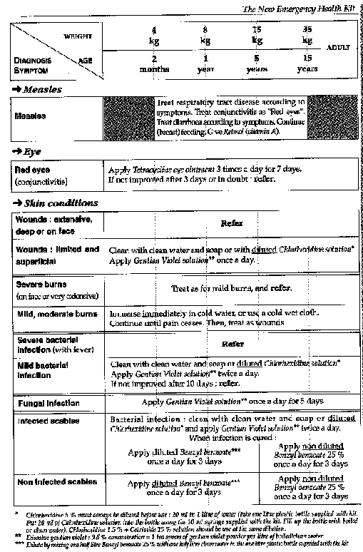
Table
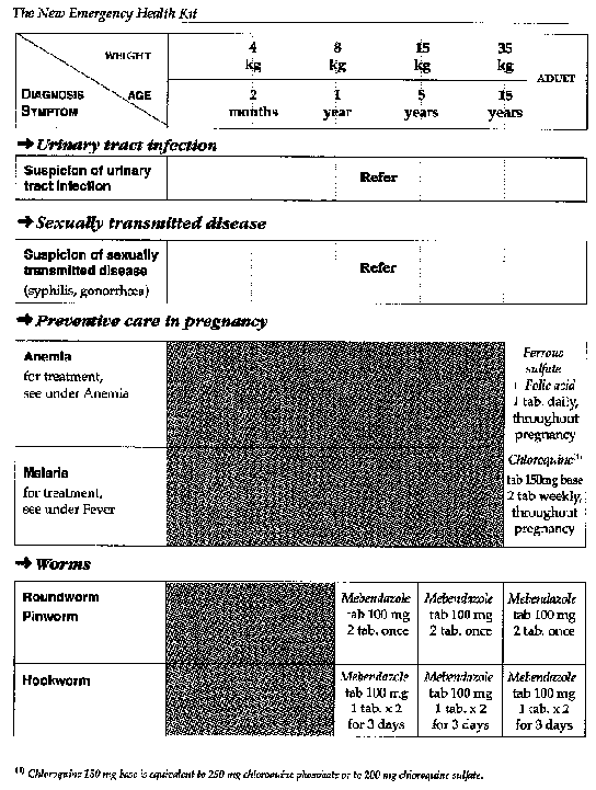
Table
Annex 2
Evaluation and treatment of diarrhoea
Assessment of diarrhoea patients for dehydration Annex 2a
Annex 2a

Table
Annex 2b
Treatment plan A to treat dirrhoea at home
Use this plan to teacb the mother to:
· Continue to treat at home her child's current episode of diarrhoea.
· Give early treatment for future episodes of diarrhoea.
Explain the tbree rules for treating diarrhoea at home
1. GIVE THE CHILD MORE FLUIDS THAN USUAL TO PREVENT DEHYDRATION:
· Use a recommended home fluid, such as a cereal gruel. If this is not possible, give plain water.
· Use ORS solution for children described in the box overleaf.
· Give as much of these fluids as the child will take. Use the amounts shown below for ORS as a guide.
· Continue giving these fluids until the diarrhoea stops.
2. GIVE THE CHILD PLENTY OF FOOD TO PREVENT UNDERNUTRITION:
· Continue to breast-feed frequently.
· If the child is not breast-fed, give the usual milk. If the child is less than 6 months old and not yet taking solid food, dilute milk of formula with an equal amount of water for 2 days.
· If the child is 6 months or older, or already taking solid food:
- Also give cereal or another starchy food mixed, if possible, with pulses, vegetables, and meat of fish. Add 1 or 2 teaspoonfuls of vegetable oil to each serving.
- Give fresh fruit juice or mashed banana to provide potassium.
- Give freshly prepared foods. Cook and mash or grind food well.
- Encourage the child to eat: offer food at least 6 times a day.
- Give the same foods after diarrhoea stops, and give an extra meal each day for two weeks.
3. TAKE THE CHILD TO THE HEALTH WORKER IF THE CHILD DOES NOT GET BETTER IN 3 DAYS OR DEVELOPS ANY OF THE FOLLOWING:
· Many watery stools
· Repeated vomiting
·
Marked thirst
· Eating or drinking poorly
· Fever
·
Blood in the stool
Children sbould be given ORS solutions at bome, if:
· They have been on Treatment Plan B or C.
· They
cannot return to the health worker if the diarrhoea gets worse.
· It is
national policy to give ORS to all children who see a health worker for
diarrhoea.
IF THE CHILD WILL BE GIVEN ORS SOLUTION AT HOME, SHOW THE MOTHER HOW MUCH ORS TO GIVE AFTER EACH LOOSE STOOL AND GIVE HER ENOUGH PACKETS FOR 2 DAYS:
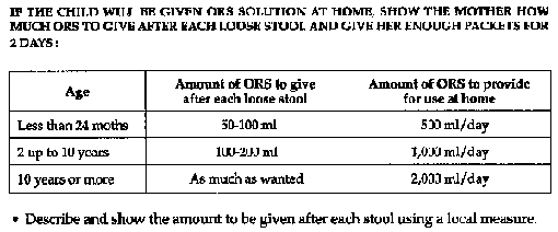
Table
· Describe and show the amount to be given after each stool using a local measure.
Show the mother bow to mix ORS.
Show her how to give ORS:
· Give a teaspoonful every 1-2 minutes for a child under 2 years.
· Give frequent sips from a cup for an older child.
· If the child vomits, wait 10 minutes. Then give the solution more slowly (for example, a spoonful every 2-3 minutes).
· If diarrhoea continues after the ORS packets are used up, tell the mother to give other fluids as described in the first rule above or return for more ORS.
Annex 2c
Treatment plan B to treat dehydration
APPROXIMATE AMOUNT OF ORS SOLUTION TO GIVE IN THE FIRST 4 HOURS:

Table
· If the child wants more ORS than shown, give more.
· Encourage the mother to continue breast-feeding.
· For infants under 6 months who are not breast-fed, also give 100-200 ml clean water during this period.
OBSERVE THE CHILD CAREFULLY AND HELP THE MOTHER GIVE ORS SOLUTION:
- Show her how much solution to give her child.
- Show her how to give it- a teaspoonful every 1-2 minutes for a child under 2 years, frequent sips from a cup for an older child.
- Check from time to time to see if there are problems.
- If the child vomits, wait 10 minutes and then continue giving ORS, but more slowly, for example, a spoonful every 2-3 minutes.
- If the child's eyelids become puffy, stop ORS and give plain water or breast milk. Give ORS according to Plan A when the puffiness is gone.
AFTER 4 HOURS, REASSESS THE CHILD USING THE ASSESSMENT CHART. THEN SELECT PLAN A, B OR C TO CONTINUE TREATMENT.
· If there are no signs of dehydration, shift to Plan A. When dehydration has been corrected, the child usually passes urine and may also be tired and fall asleep.
· If signs indicating some dehydration are still present, repeat Plan B, but start to offer food, milk and juice as described in Plan A.
· If signs indicating severe dehydration have appeared, shift to Plan C.
IF THE MOTHER MUST LEAVE BEFORE COMPLETING TREATMENT PLAN B:
· Show her how much ORS to give to finish the 4-hour treatment at home.
· Give her enough ORS packets to complete rehydation, and for 2 more days as shown in Plan A.
· Show her how to prepare ORS solution.
· Explain to her the three rules in Plan A for treating her child at home:
- to give ORS or other fluids until diarrhoea stops;
- to
feed the child;
- to bring the child back to the health worker, if necessary.
Annex 2d
Treatment plan C to treat severe dehydration quickly
Fallow the arrows. If the answer is "yes", go across. If "no", go down.

Table
Notes:
· If possible, observe the patient at least 6 hours after rehydration to be sure the mother can maintain hydration giving ORS solution by mouth.
· If the patient is above 2 years and there is cholera in your area, give an appropriate oral antibiotic after the patient is alert.
Annex 3
Management of the child with cough or difficutt breathing
· Assess the child
Ask:
-How old is the child ?
-Is the child coughing ? For how long
?
-Is the child able to drink ? (for children age 2 months up to 5
years)
-Has the child stopped feeding well ? (for children less than 2
months)
-Has the child had fever ? For how long ?
-Has the child had
convulsions ?
Look and listen (the child must be calm).
- Count the breaths in one minute.
- Look for chest
indrawing.
- Look and listen for stridor.
- Look and listen for wheeze. Is
it recurrent ?
- See if the child is abnormally sleepy, or difficult to
wake.
- Feel for fever, or low body temperature (or measure
temperature).
- Look for severe undernutrition.
· Decide how to treat the child
-The child aged less than two months see Annex 3a
-The child
aged two months up to five years see Annex 3b
- who is not wheezing
- who
is wheezing
-Treatment instructions
- Give an antibiotic
- Advise
mother to give home care see Annex 3c
- Treatment of fever
Annex 3a
The child aged less than two months

Table
Annex 3b
The child aged two months to five years

Table
Annex 3c
Treatment instructions
· Give an antibiotic
- Give first dose of antibiotic in clinic.
- Instruct mother on how to give the antibiotic for five days at home (or to retum to clinic for daily procaine penicillin injection).

Table
- Advise mother to give home care
· Feed the child.
- Feed the child during illness.
- Increase feeding after
illness.
- Clear the nose if it interferes with feeding
· Increase fluids.
- Offer the child extra to drink.
- Increase breastfeeding.
· Soothe the throat and relieve the cough with a safe remedy.
· More important: in the child classified as having "No pneumonia: cough or cold", watch for the following signs and return quickly if they occur:
- Breathing becomes difficult.
- Breathing becomes fast.
-
Child is not able to drink.
- Child becomes sicker.
This child my have pneumonia
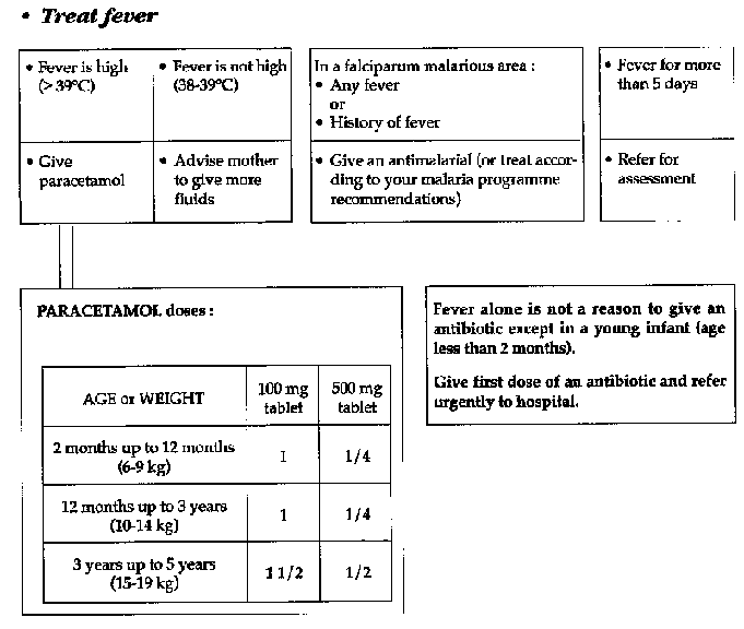
Table
Annex 4

Table
Annex 5

Table
Annex 6
Guidelines for suppliers
Quality
1. The quality of the drugs must comply with internationally recognized pharmaco poeial standards.
2. At the time of shipment the product shall have at least two thirds of its shelf life.
3. Tablets should preferably be divisible and carry characteristic symbols for easy identification.
4. Drugs should be procured only from those manufacturers able to produce documents meeting the regulations of the WHO Certification Scheme on the Quality of Pharmaceutical Products Moving in International Commerce.
Labelling
1. Labelling should be in English and preferably one other official language of WHO.
2. All labels should display at least the following information: International nonproprietary name (INN) of the active ingredient(s).
· Dosage form.
· Quantity of active ingredient(s) in the dosage form (e.g., tablet, ampoule) and the number of units per package.
· Batch number.
· Date of manufacture.
· Expiry date (in clear language, not in code).
· Pharmacopoeial standard (e.g. BP, USP...).
· Instructions for storage.
· Name and address of the manufacturer.
3. A printed label on each ampoule should contain the following:
· INN of the active ingredient(s).
· Quantity of
the active ingredient.
· Batch number.
· Name of the
manufacturer.
· Expiry date.
The full label should again appear on the collective package.
4. Directions for use, warnings and precautions may be given in leaflets (package inserts). However, such leaflets should be considered as a supplement to labelling and not as an altemative.
5. For articles requiring reconstitution prior to use (e.g. powders for injection) a suitable beyond-use time for the constituted product should be indicated.
Example of label:
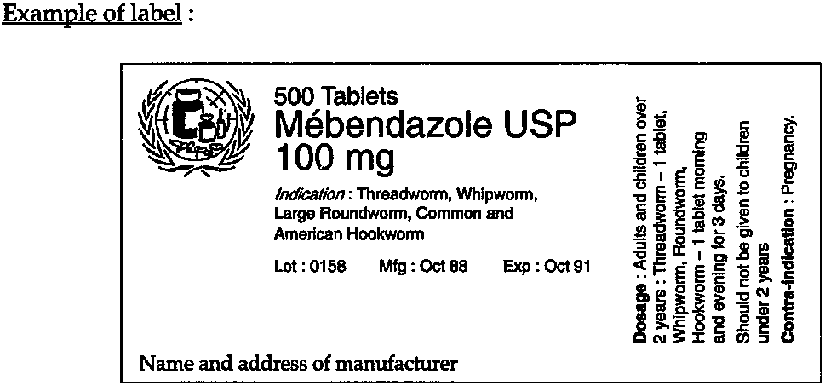
Figure
Packaging
1. Tablets and capsules should be packed in sealed waterproof containers with replaceable lid, protecting the contents against light and humidity.
2. Liquids should be packed in unbreakable leak-proof bottles or containers.
3. Containers for all pharmaceutical preparations must conform to the latest edition of internationally recognized pharmacopoeial standards.
4. Ampoules must either have break-off necks, or sufficient files must be provided.
5. Each Basic Unit should be packed in one carton. The Supplementary Unit must be packed in cartons of max. 50 kg. The cartons should preferably have two handles attached. Drugs, renewable supplies, infusions and equipment should all be packed in separate cartons, with corresponding labels.
6. Each carton must be marked with a green label (the international colour code for medical supplies in emergency situations). The word "BASIC" must be printed on each green label for the basic unit.
Packing list
Each consignment must be accompanied by a list of contents, stating the number of cartons and the type and quantity of drugs and other supplies in each carton.
Annex 7
Useful addresses
World Health Organization, Avenue Appia, CH-1211 Geneva-27, Switzerland. Telephone 41.22.7912111; telex 27821; telefax 41.22.7910746
United Nations High Commissioner for Refugees, Palais des Nations, CH-1211 Geneva-10, Switzerland. Telephone 41.22.7398111; telex 27492; telefax (general) 41.22.7319546; telefax (supplies) 7310776
UNICEF (UNIPAC), Arhusgade 129, Freeport, DK 2100, Copenhagen, Denmark. Telephone 45.31.262444; telex 19813; telefax 45.31.269421 OXFAM, 274 Branbury Road, Oxford OX2 7DZ, United Kingdom. Telephone 44.865.56777; telex 83610; telefax 44.865.57612 Medecins Sans Frontieres, 8 Rue Saint-Sabin, 75011 Paris, France. Telephone 33.1.40212929; telex 214360; telefax 33.1.48066868
International Committee of the Red Cross, 17 Avenue de la Paix, CH-1202 Geneva, Switzerland. Telephone 41.22.7346001; telex 22269; telefax 41.22.7332057
League of Red Cross and Red Crescent Societies, P.O.Box 372, CH-1211 Geneva-19, Switzerland. Telephone 41.22.7345580; telex 22555; telefax 41.22.7330395
Christian Medical Commission of the World Council of Churches, P.O.Box 66, CH-1211 Geneva-20, Switzerland. Telephone 41.22.7916111; telex 23423; telefax 41.22.791.03.61
London School of Hygiene and Tropical Medicine, Keppel Street, London WC1E 7HT, United Kingdom. Telephone 44.1.6368636; telex 8953474; telefax 44.1.4365389
International Dispensary Association, P.O.Box 3098, 1003 AB Amsterdam, The Netherlands. Telephone 31.2903.3051; telex 13566; telefax 31.2903.1854
|
| |||||||||||||||||||||||||||||||||||||||||||||||||||||||||||||||||||||||||||||||||||||||||||||||
List of medications
(commercial and common names)
Acetylsalicylic acid = A.S.A., Aspirin
Adrenaline = Epinephrine = Levorenine
Aluminium Hydroxide
Aminophylline = Theophylline = Euphyllin
Amoxycillin = Amoxil, Clamoxyl
Ampicillin = Amfipen, Penbritin
Ascorbic Acid = Vitamin C, Redoxon
Benzathine Penicillin = Benzathine Benzyl penicillin = Penidural
Benzonidazole = Benznidazole = Radanil
Benzyl Benzoate = BBL
Benzyl Penicillin = Penicillin G = Crystapen
Calcium Gluconate
Chloramine = Tosylchloramide sodium = Clonazone
Chloramphenicol = Chloromycetin, Tifomycine
Chlorhexidine + Cetrimide = HAC, Savlon
Chloroquine = Nivaquine, Resochin
Chlorpheniramine = Chlorphenamine = Teldvin
Chlorpromazine = Largactil
Cimetidine = Tagamet
Clofazimine= Lamprene
Cloxacillin = 0rbenin
Cotrimoxazole = Sulphamethoxazole + Trimethoprim = Bactrim, Cotrim, Septrim
Dapsone = Avlosulfon
Dexamethasone = Decadron, Oradexon
Diazepam = Tensium, Valium
Diethylcarbamazine = Banacide, Notezine
Digoxin = Lanoxin
Doxycycline = Doxy 100, Granudoxy, Vybramycin
Epinephrine = Adrenaline = Levorenine
Ergometrine (methyl) = Methergin
Erythromycin = Erythrocin, Ilotycin
Ethambutol = Myambutol
Etionamide = Iridocin, Trecator
Ferrous sulphate = Eryfer, Ferro Grad, Resofero
Folic Acid = Folacin, Foldine
Furosemide = Frusemide = Frusid, Lasix
Gentamicin= Cidomycin, Garamycin, Gentallin
Gentian Violet = G.V.
Griseofulvin = Fulcin, Grisovin
Hydralazine = Apresoline
Hydrochlorothiazide = Dochlotride, Esidrex, HydroSaluric
Hydrocortisone = Efcortesol, Solu-cortef
Hyoscine (N-Butyl) = Butylscopolamine = Buscopan
Indometacin = Artracin, Indocid
Isoniazid = INH = Rimifon
Ivermectin = Mectizan
Levamizol = Tramisol
Lidocaine = Lignocaine = Xylocaine, Xylocard
Mebendazole = Vermox
Mefloquine antimoniate = Methy glucamide = Glucantime
Melarsopol = Arsobal
Methyldopa = Aldomet,Medomet
Ivermectin = Mectizan
Levamizole = Tramisol
Lidocaine = Lignocaine = Xylocaine, Xylocard
Mebendazole = Vermax
Mefloquine = Lariam
Meglumine antimoniate = Methy glucamine = Glucantime
Melarsoprol = Arsobal(g)
Methyldopa = Aldomet, Medomet
Metrifonate = Bilarcil
Metronidazole = Flagyl, Metrolyl, Zadstat
Miconazole = Dactarin
Niclosamide = Tredemine, Yomesan
Nifurtimox = Lampit
Nitrofurantoin = Furandantin, Urantoin
Noramidopyrine = Dypirone = Metamizol = Nolutil, Novalgin
Norethisterone = Norlutin, Primolut
Nystatin = Mycostatin, Nystan
Oral rehydration salt = 0RS = 0ralit
Oxamniquine = Mansil, Vansil
Oxytocin= Pitocin, Syntocinon
Paracetamol = D o l i p r an, Panadolt, Tylenol
Penicillin G = Benzyl penicilline = Crystapen
Penicillin V = Phenoxymethyl penicillin = Crystapen,
Stabillin
V-K, V-Cil-K
Pentamidine = Lomidine
Pentazocine = Fortal
Phenobarbital = Phenobarbitone = Gardena, Luminal
Phenytoin = Di-Hydan, Dilantin, Epanutin
Piperazine = Antepar, Pripsen
Potassium (Chloride or Gluconate) = Kalleorid
Povidone iodine = Polyvidone iodine = Betadin, Videne
Praziquantel = Biltricide
Prednisolone = Prednisone = Codesol, Deltastab, Prednesol
Primaquine
Probenecid = Benemid
Procain Penicillin = Procain Benzyl Penicillin
Promethazine = Phenergan
Propranolol = Angilol, Inderal
Pyrantel pamoate = Combantrin
Pyrazinamide = Zinamide
Pyridoxine = Vitamin B6 = Becilan
Pyrimethamine = Daraprim, Malocide
Quinine = Quinimax, Quinoforme
Retinol = Vitamin A = Ro-A-Vit
Rifampicin = Rifadin, Rimactane
Ringer Lactate = Hartmann's solution
Salbutamol = Albuterol = Salbulin, Salbutan, Ventolin
Sodium Stibogluconate = Pentostam
Spectinomycin = Trobicin
Spironalactone = Aldacton, Osiren
Streptomycin
Sulfacetamide
Sulphadoxine + Pyrimethamine = Fansidar
Suramin sodium = Antrypol, Moranyl
Tetracycline = Abfosan, Hexacycline, Tetramig
Thiabendazole = Mintezol
Thiacetazone= TB1
Thiamine = Aneurin = Vitamin B1 = Benerva, Bevitine
Trimethoprime + Sulphamethoxazole = Cotrimoxazole = Bactrim, Cotrim, Septrim
Vitamin A = Retinol = Ro-A-Vit
Whitfield's ointment = 3 % Salycilic Acid + 6 % Benzoic Acid
|
| |||||||||||||||||||||||||||||||||||||||||||||||||||||||||||||||||||||||||||||||||||||||||||||||
Bibliography
1. AGREGES DU PHARO
Techniques elementaires pour medecins
isoles
DGDL Editions, 1983
2. ALLEGRA D. et Col.
Emergency Refugee Health Care: A
chronicle of experience in Khmer assistance operation 1979-1980
Centers for
Diseases Control, Atlanta, 1983
3. AMERICAN PUBLIC HEALTH ASSOCIATION
Control of communicable
diseases in man
Abram S. Benenson Editeur, 1985
4. BERKOW R.
The Merck manuel
Merck Sharp and Dohme, 1987
5. BRIEND A.
Prevention et traitement de la
malnutrition
Orstom Editions, 1985
6. BRITISH MEDICAL ASSOCIATION
British National
Formulary
British Medical Association and the Pharmaceutical Society of Great
Britain, 1987
7. CAMERON M., HOFVANDER Y.
Manual on feeding infants and
young children
Orstom, 1983
8. DAWSON C.R., JONES B.R., TARIZZO M.L.
Guide pour la lutte
contre le trachome
W.H.O., Geneva, 1981
9. FERRIER E.
Precis de Pediatrie
Payot Edit., 1982
10. GENTILINI M., DUFLO B.
Medecine Tropicale
Flammarion
Sciences, 1986
11. JELLIFE D.B., STANFELD P.l.
Diseases of Children in the
Subtropics and Tropics
Edward Arnold, 1978
12. H.C.R.
Manuel des situations d'urgence
H.C.R., Geneva,
1982
13. HAROLD FRIEDMAN H.
Manuel de diagnostic medical
Mesdi
Editions, 1984
14. HERCBERG S. et Col.
Nutrition et Sante
Publique
Lavoisier Paris, 1986
15. HUAULT G., LABRUNE B.
Pediatrie d'urgence
Flammarion
Medecine Sciences, 1981
16. KENNETH R., NISWANDER
Manuel d'obstetrique
Mesdi
Edition, 1985
17. KERNBAUM S.
Elements de Pathologie Infectieuse
Simep -
Specia, 1982
18. KING M.
Primary Child Care
Oxford Medical
Publications, 1983
19. Malawi Ministry of Health
Guideline for management of
malaria
Ministry of Health, Malawi, 1986
20. MANSON BAHR
Manson's tropical diseases
Bailliere and
Tindall Edition, 1982
21. MARTINDALE
The extra pharmacopoeia
The Pharmaceutical
press, London, 1982
22. MEDECINS SANS FRONTIERES
Essential drugs - practical
guidelines
Editions Hatier, Paris, 1993
23. MEDECINS SANS FRONTIERES
Principales conduites a tenir en
dispensaire - Niveau auxiliaire medical
Medecins sans Frontieres, Paris, 1990
24. MORLEY D.
Pediatrie dans les pays en voie de
developpement
Flammarion Medecine Sciences 1977
25. O.M.S./W.H.O.
Fievre hemorragique virale
Technical
report n°721, OMS, Geneva, 1985
26. O.M.S./W.H.O.
La lutte contre la carence en Vitamine A et
la Xerophtalmie
Technical report n°672, OMS, Geneva, 1982
27. O.M.S./W.H.O.
La situation du paludisme dans le monde en
1990, parties I et II
Releve epidemiologique hebdomadaire, 67 (22/23)
WHO,
Geneva, 1992
28. O.M.S./W.H.O.
Voyages intemationaux et sante -
Vaccinations et conseils d´hygiene: situation au 01/01/91
WHO, Geneva,
1991
29. O.M.S./W.H.O.
Manuel pour la lutte contre la
trypanosomiase
WHO, Geneva, 1983
30. O.M.S./W.H.O.
Comite d'experts de la rage - Rapports
techniques N°709
WHO, Geneva, 1984
31. O.M.S./W.H.O.
Nutrition in preventive medicine
WHO,
Geneva, 1976
32. O.M.S./W.H.O.
L'utilisation des medicaments
essentials
Technical report n° 722 - WHO, Geneva, 1985
33. O.M.S./W.H.O.
La lutte contre les maladies sexuellement
transmissibles
WHO, Geneva, 1986
34. O.M.S./W.H.O.
Prevention and Control of Yellow Fever in
Africa
WHO, Geneva, 1986
35. O.M.S./W.H.O.
Traitement et prevention des diarrhees
aigues: directives destinees aux instructeurs des agents de sante
WHO,
Geneva, 1985
36. O.M.S./W.H.O.
Programme for the control of diarrhoeal
disease - a manual for the treatment of diarrhoea
WHO/CDD/SER/80.2 REV. 2,
Geneva, 1990
37. O.M.S. /W.H.O.
Programme de lutte contre les maladies
diarrheiques: directives pour une enquete par sondage sur les taux de morbidite,
de mortalite et de traitement relatifs aux maladies
diarrheiques
OMS/LMD/SER/84.6 REVI, Geneva, 1984
38. O.M.S./W.H.O.
Respiratory infections in children:
management at small hospitals.
Background notes and a manual for
doctors
WHO, Geneva, 1986
39. OXFAM
Guide pratique de programmes selectifs
d'alimentation
OXFAM, 1984
40. OXFAM
Guidelines for tuberculosis control programmes in
developing countries
OXFAM, 1985
41. PECHERE J.C. et Col.
Les infections
EDISEM, Maloine,
Paris, 1985
42. PENE P., ANDRE JE, ROUGEMONT A.
Sante et Medecine en
Afrique Tropicale
Doin Edition, 1980
43. PERELMAN R.
Pediatrie pratique
Maloine Editeur, 1977
44. PERINE P.L. et Col.
Manuel des treponematoses
endemiques
OMS, Geneve, 1985
45. PERRIN Pierre
Assistance medicale en situation
d'urgence
C.I.C.R., 1984
46. PETERSDORF, ADAMS, BRAUNWALD, ISSELBACHER, MARTIN,
WILSON
Harrison's principles of internal medicine
Internal student
association, 1983
47. REESE R.E. et DOUGLAS R.G.
A practical approach to
infectious diseases
Little Brown and Company, Boston/Toronto, 1986
48. ROTSART, HERTHING I., COURTEJOIE J.
Maternite et
Sante
Bureau d'Etudes et de Recherches pour la promotion de Sante Zaire, 1983
49. SIMMONDS S. et Col.
Refugee community health care
Ross
Oxford Medical publications, 1983
50. UNICEF
Assisting in emergencies
UNICEF, New York, 1986
51. WARREN K.S., MAHMOUD A.F.
Tropical and geographical
medicine
Mc Graw Hill Book Company, New York, 1984
FRANCE Medecins Sans Frontieres 8 rue Saint-Sabin- 75544 Paris Cedex 11 Tel.: (33) 1- 40.21.29.29 - Fax: (33) 1- 48.06.68.68 Telex (042) 214360 MSF F
BELGIUM Medecins Sans Frontieres 24 rue Deschampheleer - 1080 Bruxelles Tel.: (32) 2- 414.03.00 - Fax: (32) 2- 411.82.60 Telex: (046) 63607 MSF B
NETHERLANDS Artsen Zonder Grenzen Postadres - Postbus 10014- 1001 EA Amsterdam Tel.: (31) 20- 520.87.00 - Fax: (31) 20- 620.51.70 Telex: (044) 10773 MSF NL
SWITZERLAND Medecins Sans Frontieres 3 Clos de la Fonderie - 1227 Carouge / Geneve Tel.: (41) 22- 300.44.45 - Fax: (41) 22- 300.44.14 Telex: (045) 421927 MSF CH
SPAIN Medicos Sin Fronteras Avenida Portal del Angel, n°l, 1 - 08002 Barcelona Tel.: (34) 3- 412.52.52 - Fax: (34) 3- 302.28.89 Telex: (052) 97309 MSF E
LUXEMBOURG Medecins Sans Frontieres 70 route de Luxembourg - L-7240 Bereldange Tel.: (352) 33.25.15 - Fax: (352) 33.51.33 Telex: (0402) 60811 MSF LU
GREECE Giatri Horis Synora 11 A. Paioniou - 10440 Athenes Tel.: (30) 1- 88.35.334 - Fax: (30) 1- 88.29.988
Imprime en France par ISI, 75011 Paris
Deput legal
n°9288 - janvier
1994